File:Stage 22 image 217.jpg: Difference between revisions
mNo edit summary |
|||
| Line 32: | Line 32: | ||
{{Carnegie_stage_table_1}} | {{Carnegie_stage_table_1}} | ||
{{Neural Table}} | |||
<references/> | <references/> | ||
Revision as of 17:32, 26 May 2017
Human Embryo Developing Cortex
The ventricle is the CSF fluid-filled space within the brain. The inset image shows lying within the ventricles, part of the choroid plexus that forms and secretes the CSF. Outside the developing cortex the developing brain meninges pia and sub-arachnoid layers can also be seen. |

Low resolution image |
- Links: Image - cortex overview | Image - cortex layers | Image - high resolution version | Cerebrum Development | Meninges Development
Note this section image is vertically flipped compared with original serial section images.
| Selected Embryo Histology - Week 8 (Stage 22) |
|---|
|
| Links: Carnegie stage 22 | Week 8 |


| Stage 22 Links: Week 8 | System Development | Lecture - Limb | Lecture - Head Development | Lecture - Sensory | Science Practical - Head | Science Practical - Sensory | Science Practical - Urogenital | Historic - Skull Development | Carnegie Embryos | Madrid Embryos | Category:Carnegie Stage 22 | Next Stage 23 |
| Week 8, GA week 10, 54 - 56 days, CRL 23 - 28 mm, Carnegie Embryos |
| Historic Papers: 1914 | 1954 Stage 19-23 |
- Carnegie Stages: 1 | 2 | 3 | 4 | 5 | 6 | 7 | 8 | 9 | 10 | 11 | 12 | 13 | 14 | 15 | 16 | 17 | 18 | 19 | 20 | 21 | 22 | 23 | About Stages | Timeline
| Week: | 1 | 2 | 3 | 4 | 5 | 6 | 7 | 8 |
| Carnegie stage: | 1 2 3 4 | 5 6 | 7 8 9 | 10 11 12 13 | 14 15 | 16 17 | 18 19 | 20 21 22 23 |
| Neural Tube | Primary Vesicles | Secondary Vesicles | Adult Structures |
|---|---|---|---|
| week 3 | week 4 | week 5 | adult |
| prosencephalon (forebrain) | telencephalon | Rhinencephalon, Amygdala, hippocampus, cerebrum (cortex), hypothalamus, pituitary | Basal Ganglia, lateral ventricles | |
| diencephalon | epithalamus, thalamus, Subthalamus, pineal, posterior commissure, pretectum, third ventricle | ||
| mesencephalon (midbrain) | mesencephalon | tectum, Cerebral peduncle, cerebral aqueduct, pons | |
| rhombencephalon (hindbrain) | metencephalon | cerebellum | |
| myelencephalon | medulla oblongata, isthmus | ||
| spinal cord, pyramidal decussation, central canal | |||
- ↑ <pubmed>5414696</pubmed>
Cite this page: Hill, M.A. (2024, April 19) Embryology Stage 22 image 217.jpg. Retrieved from https://embryology.med.unsw.edu.au/embryology/index.php/File:Stage_22_image_217.jpg
- © Dr Mark Hill 2024, UNSW Embryology ISBN: 978 0 7334 2609 4 - UNSW CRICOS Provider Code No. 00098G
File history
Click on a date/time to view the file as it appeared at that time.
| Date/Time | Thumbnail | Dimensions | User | Comment | |
|---|---|---|---|---|---|
| current | 01:09, 23 August 2011 | 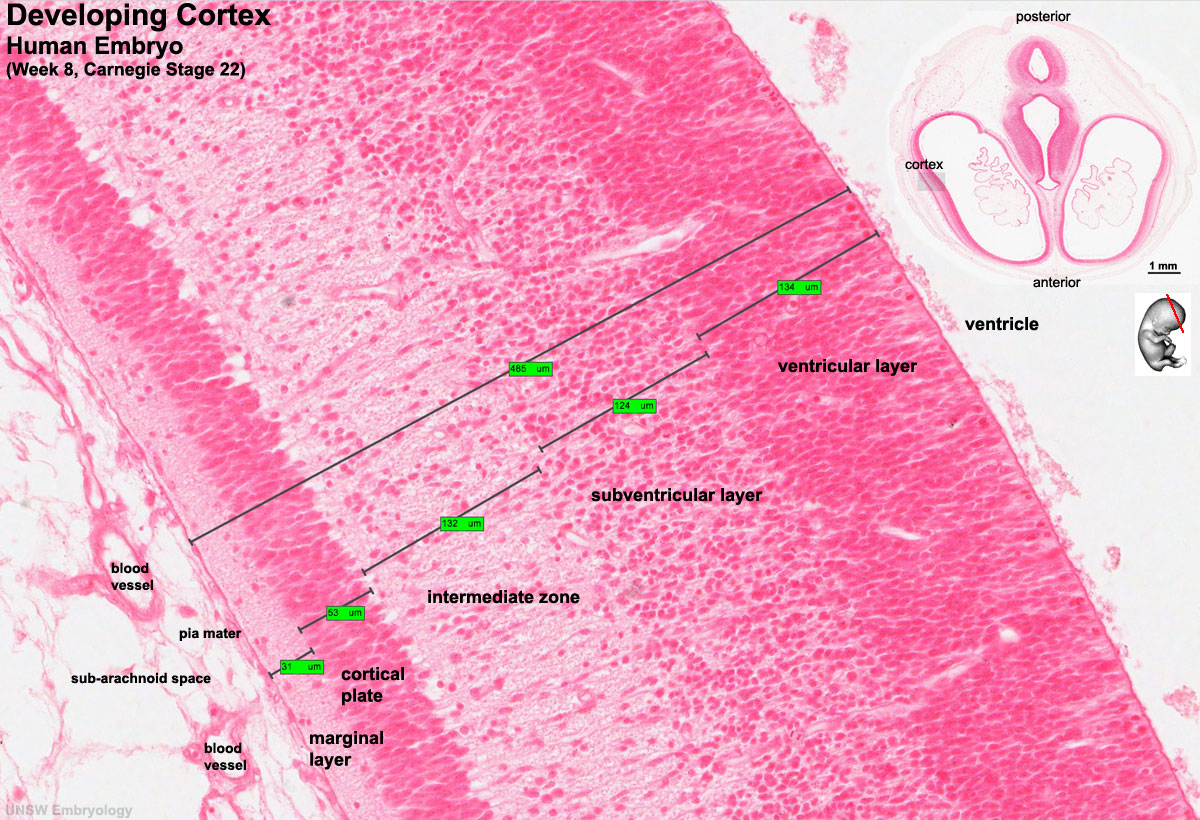 | 1,200 × 820 (323 KB) | S8600021 (talk | contribs) | ==Human Embryo Developing Cortex== * Week 8, Carnegie stage 22 * Note this section image is vertically flipped compared with serial original serial images. :Links: Neural System Development Cortex-with-section-insert-4.jpg (scan 35815 cropped and sc |
You cannot overwrite this file.
File usage
The following 50 pages use this file:
- 2017 Group Project 1
- AE Practical - Neural Histology
- ANAT2241 Nervous Tissue
- ANAT3411 Neuroanatomy
- BGDA Lecture - Development of the Embryo/Fetus 2
- BGDA Lecture - Development of the Nervous System
- BGDA Practical 12 - Embryo to Fetus
- BGDA Practical 7 - Week 8
- Brain Awareness Week 2012
- Carnegie stage 22
- K12 Brain Awareness Week
- Lecture - Neural Development
- Neural - Cerebrum Development
- Neural - Meninges Development
- Neural System Development
- Talk:2017 Group Project 1
- Talk:ANAT3411 Neuroanatomy
- User:Maria Angeles
- File:Stage22 HPA2L.jpg
- File:Stage22 vertebra and spinal cord 1.jpg
- File:Stage 22 image 200.jpg
- File:Stage 22 image 201.jpg
- File:Stage 22 image 203.jpg
- File:Stage 22 image 204.jpg
- File:Stage 22 image 205.jpg
- File:Stage 22 image 206.jpg
- File:Stage 22 image 207.jpg
- File:Stage 22 image 208.jpg
- File:Stage 22 image 209.jpg
- File:Stage 22 image 210.jpg
- File:Stage 22 image 211.jpg
- File:Stage 22 image 212.jpg
- File:Stage 22 image 213.jpg
- File:Stage 22 image 214.jpg
- File:Stage 22 image 215.jpg
- File:Stage 22 image 216.jpg
- File:Stage 22 image 217.jpg
- File:Stage 22 image 218.jpg
- File:Stage 22 image 219.jpg
- File:Stage 22 image 220.jpg
- File:Stage 22 image 222.jpg
- File:Stage 22 image 223.jpg
- File:Stage 22 image 224.jpg
- File:Stage 22 image 225.jpg
- File:Stage 22 image 301.jpg
- File:Stage 22 image 302.jpg
- File:Stage 22 image 322.jpg
- File:Stage 22 vomeronasal organ.jpg
- Template:Stage 22 histology gallery
- Template:Stage 22 histology gallery table