File:Stage 13 image 101.jpg: Difference between revisions
({| |-bgcolor="DDCEF2" | width=400px| :'''Links:''' G7L | Unlabeled | Previous | Last Image | All sections |} ===Cardiovascular Syste) |
No edit summary |
||
| Line 2: | Line 2: | ||
|-bgcolor="DDCEF2" | |-bgcolor="DDCEF2" | ||
| width=400px| | | width=400px| | ||
:'''Links:''' G7L | [[:File:Stage 13 image 049.jpg|Unlabeled]] | [[:File:Stage 13 image | :'''Links:''' G7L | [[:File:Stage 13 image 049.jpg|Unlabeled]] | [[:File:Stage 13 image 098.jpg|Labeled]] | [[Carnegie_stage_13_-_serial_sections#Labeled_Sections|All sections]] | ||
|} | |} | ||
===Cardiovascular System=== | ===Cardiovascular System=== | ||
Revision as of 09:20, 16 December 2010
|
Cardiovascular System
Cardiovascular System Development
- Heart showing both ventricles (and ventricular septum) and a single atria in this section.
- Aortic sac lying ventral to the hypopharyngeal eminence.
- Ductus venosus large cavity within the liver.
- Truncus arteriosus above heart.
- Dorsal aorta with intersegmental artery branches lying ventral to developing vertebral column.
- Caudal pharynx (extending laterally, ventral to dorsal aorta - compare with B4).
- Stomach and lesser sac in mesentery.
- Triangular flange of mesentery with triangular hole and intervitelline anastomosis.
- Hindgut (without lumen) seen at caudal end of mesentery.
- dorsal aorta with its dorsal segmental arterial branches.
- The dorsal segmental artery itself marks the location of the centre of the light-staining part of the sclerotome, which is the future vertebral body.
Original File name: PigG7L.jpg http://embryology.med.unsw.edu.au/wwwpig/pigg/G7L.htm
| System Links: Introduction | Cardiovascular | Coelomic Cavity | Endocrine | Gastrointestinal Tract | Genital | Head | Immune | Integumentary | Musculoskeletal | Neural | Neural Crest | Placenta | Renal | Respiratory | Sensory | Birth |
About Stage 13 Embryo Sections - This image is from a serial section of a 6mm CRL pig embryo with some features of the Stage 14 embryo. This embryo is approximately equal to the day 42 human embryo. Use these serial images to identify internal features and relationships that exist within the embryo at this stage. Then compare these images with the later features of the Carnegie stage 22 human embryo.
| Stage 13 Serial unlabeled images | Embryo Stage 13 Serial labeled images |
Cite this page: Hill, M.A. (2024, April 19) Embryology Stage 13 image 101.jpg. Retrieved from https://embryology.med.unsw.edu.au/embryology/index.php/File:Stage_13_image_101.jpg
- © Dr Mark Hill 2024, UNSW Embryology ISBN: 978 0 7334 2609 4 - UNSW CRICOS Provider Code No. 00098G
File history
Click on a date/time to view the file as it appeared at that time.
| Date/Time | Thumbnail | Dimensions | User | Comment | |
|---|---|---|---|---|---|
| current | 09:20, 16 December 2010 | 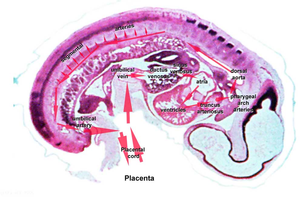 | 1,000 × 649 (65 KB) | S8600021 (talk | contribs) | {| |-bgcolor="DDCEF2" | width=400px| :'''Links:''' G7L | Unlabeled | Previous | Last Image | All sections |} ===Cardiovascular Syste |
You cannot overwrite this file.
File usage
The following page uses this file: