File:Stage 13 image 097.jpg
Original file (1,000 × 720 pixels, file size: 162 KB, MIME type: image/jpeg)
G6L Image Features
|
Gastrointestinal Development
Gastrointestinal Tract Development
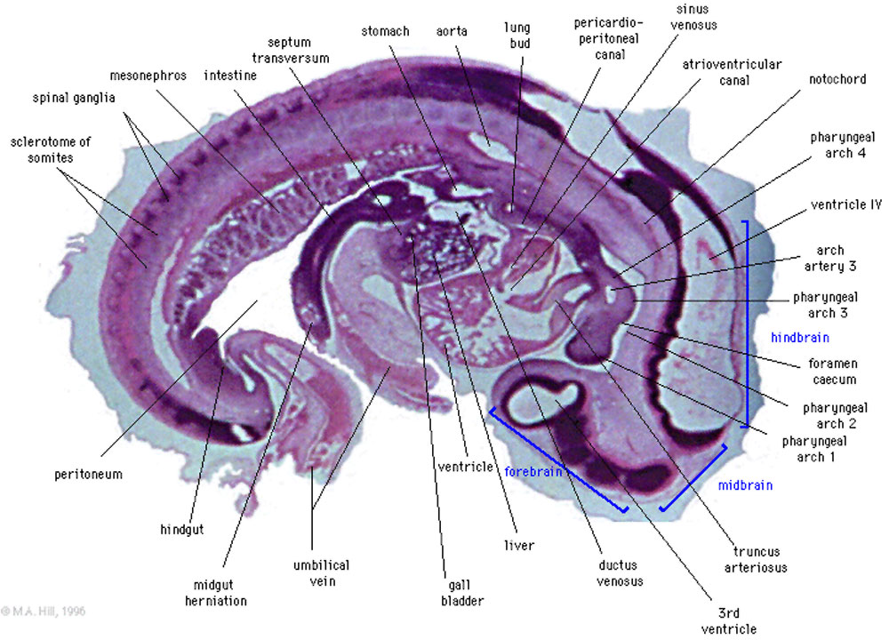
- Rathke's pouch. Floor of pharynx with foramen caecum (remains of thyroglossal duct), and caudally to it, the hypopharyngeal eminence.
- Aortic sac ventral to H-P eminence.
- Caudal pharynx (extending laterally, ventral to dorsal aorta - cf B4). L lungbud caudal to L atrium with attachment of pulmonary mesenchyme to septum transversum.
- Stomach, mesentery.
- Liver. Gall bladder.
- Cranial limb of midgut loop in mesentery with intervitelline anastomoses.
- Caudal attachment of mesentery with hindgut inside.
- Communication of intra- and extra-embryonic coeloms.
Cardiovascular System
Cardiovascular System Development
Musculoskeletal System
Musculoskeletal System Development
- Cervical region (neural tube cut very obliquely): note the wavy notochord and thin roof and floor plates.
- Thoracic region: alternating light and dark parts of the sclerotomes (vertebral bodies and I.V. Discs, respectively).
- Lumbar region: tiny, dorsal segmental branches of dorsal aorta. Each little branch is aligned almost parallel to the cranial border of the next caudal dorsal root ganglion (segmented dark masses).
- Sacral region: oblique section of neural tube, notochord.
Renal System
About Stage 13 Embryo Sections - This image is from a serial section of a 6mm CRL pig embryo with some features of the Stage 14 embryo. This embryo is approximately equal to the day 42 human embryo. Use these serial images to identify internal features and relationships that exist within the embryo at this stage. Then compare these images with the later features of the Carnegie stage 22 human embryo.
| Stage 13 Serial unlabeled images | Embryo Stage 13 Serial labeled images |
| System Links: Introduction | Cardiovascular | Coelomic Cavity | Endocrine | Gastrointestinal Tract | Genital | Head | Immune | Integumentary | Musculoskeletal | Neural | Neural Crest | Placenta | Renal | Respiratory | Sensory | Birth |
Cite this page: Hill, M.A. (2024, April 24) Embryology Stage 13 image 097.jpg. Retrieved from https://embryology.med.unsw.edu.au/embryology/index.php/File:Stage_13_image_097.jpg
- © Dr Mark Hill 2024, UNSW Embryology ISBN: 978 0 7334 2609 4 - UNSW CRICOS Provider Code No. 00098G
File history
Click on a date/time to view the file as it appeared at that time.
| Date/Time | Thumbnail | Dimensions | User | Comment | |
|---|---|---|---|---|---|
| current | 17:24, 10 August 2010 | | 1,000 × 720 (162 KB) | S8600021 (talk | contribs) | ==Stage 13== ==Image Features== Rathke's pouch. Floor of pharynx with foramen caecum (remains of thyroglossal duct), and caudally to it, the hypopharyngeal eminence. Aortic sac ventral to H-P eminence. Caudal pharynx (extending laterally, ventral to dors |
You cannot overwrite this file.
File usage
The following 23 pages use this file:
- 2010 Lab 5
- 2010 Lab 8
- 2011 Lab 5 - Early Embryo
- ANAT2341 Lab 3 - Week 4
- ANAT2341 Lab 5 - Early Embryo
- ANAT3411 Neuroanatomy
- BGDB Gastrointestinal - Early Embryo
- Carnegie stage 13
- Carnegie stage 13 - serial sections
- Embryo Serial Sections
- Fetal ECHO Meeting 2012
- Gastrointestinal Tract - Carnegie Stage 13
- Gastrointestinal Tract - Liver Development
- Museum of Natural History Berlin - 2013 Seminar
- Neural 3D stage 13 Movie
- Placenta - Stage 13
- RPAH Cardiac Embryology 2014
- Renal System - Carnegie Stage 13
- Respiratory System - Carnegie Stage 13
- Talk:Carnegie stage 13 - serial sections
- Talk:Renal System - Carnegie Stage 13
- Template:Stage13Licon120
- Template talk:Stage13Licon120