File:Stage 11 historic-Atwell1930-4.jpg
Original file (1,000 × 1,121 pixels, file size: 114 KB, MIME type: image/jpeg)
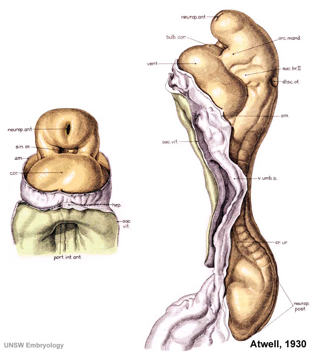
Human Embryo Carnegie stage 11
Historic drawing of the Carnegie stage 11, 19 somite pairs, approx 25 days. Ventral and Lateral external views, amnion and yolk sac removed.
- Left hand image - ventral view, rostral half of embryo, neural tube, anterior neuropore, posterior neuropore, cardiac bulge, liver, midgut opening
- Right hand image - lateral view, whole embryo, neural tube, anterior neuropore, cardiac bulge, otic placode, somites
About Carnegie stage 11
- day 23 to 26
- size 2.5 - 4.5mm CRL
- somite pairs number 13 - 20
Features: heart, neural tube, forebrain, midbrain, hindbrain, posterior neuropore, somites, sensory placodes
Original Image name: Historic-Atwell-1930-slide65.jpg, Stage 11 historic-Atwell1930-4.jpg
- Image links: External view 17 somite pairs | Sagittal section | upper embryo | lower embryo | Carnegie stage 11
Cite this page: Hill, M.A. (2024, April 19) Embryology Stage 11 historic-Atwell1930-4.jpg. Retrieved from https://embryology.med.unsw.edu.au/embryology/index.php/File:Stage_11_historic-Atwell1930-4.jpg
- © Dr Mark Hill 2024, UNSW Embryology ISBN: 978 0 7334 2609 4 - UNSW CRICOS Provider Code No. 00098G
File history
Click on a date/time to view the file as it appeared at that time.
| Date/Time | Thumbnail | Dimensions | User | Comment | |
|---|---|---|---|---|---|
| current | 16:54, 3 September 2009 | | 1,000 × 1,121 (114 KB) | S8600021 (talk | contribs) | Human Embryo Historic drawing of the Carnegie stage 11, 20 somite pairs, approx 25 days. Lateral sectional view, rostral half, amnion and yolk sac removed :Left hand image - showing neural tube, gastrointestinal tract, pericardial cavity, connecting s |
You cannot overwrite this file.
File usage
The following 2 pages use this file: