File:Stage9 sem4c.jpg
From Embryology
No higher resolution available.
Stage9_sem4c.jpg (321 × 400 pixels, file size: 19 KB, MIME type: image/jpeg)
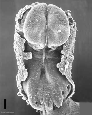
Human Embryo Carnegie Stage 9
Stage 9 day 20, somites 3-4 (scale bar 100 μm)
Dorsal view (amniotic sac cut away) showing the embryo and cut amnion edge.
Note:
- the shape of the early folded embryonic disc and rostro-caudal bendings.
- the neural groove and brain fold.
- the earliest forming somites.
- the connecting stalk.
- Carnegie Stages: 1 | 2 | 3 | 4 | 5 | 6 | 7 | 8 | 9 | 10 | 11 | 12 | 13 | 14 | 15 | 16 | 17 | 18 | 19 | 20 | 21 | 22 | 23 | About Stages | Timeline
Template:Carnegie stages table1 Image Source: Scanning electron micrographs of the Carnegie stages of the early human embryos are reproduced with the permission of Prof Kathy Sulik, from embryos collected by Dr. Vekemans and Tania Attié-Bitach. Images are for educational purposes only and cannot be reproduced electronically or in writing without permission.
File history
Click on a date/time to view the file as it appeared at that time.
| Date/Time | Thumbnail | Dimensions | User | Comment | |
|---|---|---|---|---|---|
| current | 17:23, 22 August 2009 | | 321 × 400 (19 KB) | S8600021 (talk | contribs) |
You cannot overwrite this file.
File usage
The following 28 pages use this file:
- 2010 BGD Lecture - Development of the Embryo/Fetus 2
- 2010 BGD Practical 3 - Week 3 Summary
- 2010 BGD Practical 6 - Week 3
- 2010 BGD Practical 6 - Week 8
- 2010 BGD Tutorial - Applied Embryology and Teratology
- 2010 Lab 2
- 2010 Lab 3
- 2010 Lecture 6
- 2011 Group Project 11
- Abnormal Development - Environmental
- Abnormal Development - Illegal Drugs
- Abnormal Development - Teratogens
- BGDA Lecture - Development of the Embryo/Fetus 2
- BGDA Practical 3 - Week 3 Summary
- BGDA Practical 7 - Week 3
- BGDA Practical 7 - Week 8
- BGD Tutorial - Applied Embryology and Teratology
- Carnegie stage 9
- Foundations Practical - Critical Periods
- Human Abnormal Development
- Human Embryo SEM
- Lecture - Ectoderm Development
- Lecture - Fetal Development
- Scanning Electron Microscopy
- Talk:2011 Lab 3
- File:Human-critical periods of development.jpg
- File talk:Human-critical periods.jpg
- Template:Critical Periods table