File:Stage9 sem3c.jpg: Difference between revisions
No edit summary |
No edit summary |
||
| (2 intermediate revisions by the same user not shown) | |||
| Line 1: | Line 1: | ||
== Human embryo == | == Human embryo (Carnegie Stage 9) Notochordal Plate== | ||
Stage 9 day 20, somites 3-4 | Stage 9 day 20, somites 3-4 | ||
| Line 5: | Line 5: | ||
Scanning EM selected region showing notochordal plate running rostro-caudal (right to left) from the [[:File:Stage9 sem2.jpg|ventrolateral view ]] (cut away from amniotic sac and yolk sac) showing the embryo and connecting stalk. | Scanning EM selected region showing notochordal plate running rostro-caudal (right to left) from the [[:File:Stage9 sem2.jpg|ventrolateral view ]] (cut away from amniotic sac and yolk sac) showing the embryo and connecting stalk. | ||
[[:File:Stage9 sem2.jpg|ventrolateral view ]] is a more ventral and rostral view of [[:File:Stage9 sem1.jpg|Stage9 sem1]] image. | |||
'''Links:''' [[:File:Stage9 sem2.jpg|ventrolateral view ]] is a more ventral and rostral view of [[:File:Stage9 sem1.jpg|Stage9 sem1]] image. | |||
Note: | Note: | ||
| Line 19: | Line 20: | ||
{{Template:SEM}} | {{Template:SEM}} | ||
[[Category:Human]] [[Category:Carnegie Stage 9]] [[Category:Week 3]] | |||
Latest revision as of 08:31, 8 August 2011
Human embryo (Carnegie Stage 9) Notochordal Plate
Stage 9 day 20, somites 3-4
Scanning EM selected region showing notochordal plate running rostro-caudal (right to left) from the ventrolateral view (cut away from amniotic sac and yolk sac) showing the embryo and connecting stalk.
Links: ventrolateral view is a more ventral and rostral view of Stage9 sem1 image.
Note:
- the notochordal plate on ventral surface of embryo.
- the large cilia present on the notochordal plate region.
- the numerous small villi present on the entire epithelial cell layer.
- large pale "blob" in centre of image is an artifact.
Original file name: Stage9day20somites3-4lateralsem3-600px.jpg
- Carnegie Stages: 1 | 2 | 3 | 4 | 5 | 6 | 7 | 8 | 9 | 10 | 11 | 12 | 13 | 14 | 15 | 16 | 17 | 18 | 19 | 20 | 21 | 22 | 23 | About Stages | Timeline
Image Source: Scanning electron micrographs of the Carnegie stages of the early human embryos are reproduced with the permission of Prof Kathy Sulik, from embryos collected by Dr. Vekemans and Tania Attié-Bitach. Images are for educational purposes only and cannot be reproduced electronically or in writing without permission.
File history
Click on a date/time to view the file as it appeared at that time.
| Date/Time | Thumbnail | Dimensions | User | Comment | |
|---|---|---|---|---|---|
| current | 15:02, 22 August 2009 | 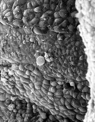 | 313 × 400 (25 KB) | S8600021 (talk | contribs) | Stage9day20somites3-4lateralsem3-600px.jpg |
You cannot overwrite this file.
File usage
The following 2 pages use this file: