File:Stage9 sem1b.jpg
Stage9_sem1b.jpg (600 × 377 pixels, file size: 33 KB, MIME type: image/jpeg)
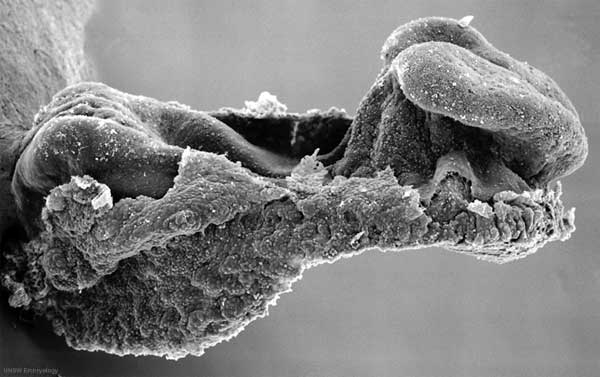
Human embryo
Stage 9, day 20, somites 3-4
Scanning EM lateral view (cut away from amniotic sac and yolk sac) showing the embryo and connecting stalk.
Note:
- the relative size of the embryo.
- the shape of the early folded embryonic disc and rostro-caudal bendings.
- the size and shape of the brain fold region.
- the primitive node and streak (left) beside connecting stalk.
- the position of the prechordal plate (cardiogenic region) ventral to brain fold.
Original file name: Stage9day20somites3-4lateralsem1-1000px.jpg
Image version links
Large 1000px | 800px | Medium 600px | Small 400px
Image Source: Scanning electron micrographs of the Carnegie stages of the early human embryos are reproduced with the permission of Prof Kathy Sulik, from embryos collected by Dr. Vekemans and Tania Attié-Bitach. Images are for educational purposes only and cannot be reproduced electronically or in writing without permission.
File history
Click on a date/time to view the file as it appeared at that time.
| Date/Time | Thumbnail | Dimensions | User | Comment | |
|---|---|---|---|---|---|
| current | 12:26, 22 August 2009 | | 600 × 377 (33 KB) | S8600021 (talk | contribs) | Human embryo Stage 9 day 20, somites 3-4 Scanning EM lateral view (cut away from amniotic sac and yolk sac) showing the embryo and connecting stalk. Note: # the relative size of the embryo. # the shape of the early folded embryonic disc and rostro-cau |
You cannot overwrite this file.
File usage
The following 10 pages use this file:
- 2010 BGD Practical 3 - Week 3 Summary
- 2010 Lab 2
- BGDA Lecture - Development of the Embryo/Fetus 2
- BGDA Practical 3 - Week 3 Summary
- Human Embryo SEM
- Timeline human development
- Talk:Timeline human development
- Template:First Trimester Timeline
- Template:First Trimester Timeline collapsable table
- Template talk:First Trimester Timeline