File:Stage9 bf3.jpg: Difference between revisions
No edit summary |
|||
| (One intermediate revision by the same user not shown) | |||
| Line 1: | Line 1: | ||
== Human | ==Human Embryo - Carnegie Stage 9== | ||
Stage 9 day 20, somites 3-4 | Stage 9 day 20, somites 3-4 | ||
Ventrolateral view (cut away from chorion and amniotic sac) showing the embryo and yolk sac. | Ventrolateral view (cut away from chorion and amniotic sac) showing the embryo and {{yolk sac}}. The first {{somite}} pairs are visible. | ||
Note: | Note: | ||
| Line 12: | Line 12: | ||
# the position of the prechordal plate (cardiogenic region) ventral to brain fold. | # the position of the prechordal plate (cardiogenic region) ventral to brain fold. | ||
{{Carnegie Stage 9 image links}} | |||
{{ | |||
Latest revision as of 23:22, 29 October 2018
Human Embryo - Carnegie Stage 9
Stage 9 day 20, somites 3-4
Ventrolateral view (cut away from chorion and amniotic sac) showing the embryo and yolk sac. The first somite pairs are visible.
Note:
- the relative size of the embryo and the associated extra-embryonic coeloms.
- the shape of the early folded embryonic disc and rostro-caudal bendings.
- the relative thicknesses of the embryo and extra-embryonic membranes.
- the position of the prechordal plate (cardiogenic region) ventral to brain fold.
Carnegie Stage 9 Image Links: (below are listed image size versions of each image)
- Light Image dorsal - Large 1000px | 800px | Medium 600px | Small 400px
- Light Image ventrolateral - Large 1000px | 800px | Medium 600px | Small 400px
- Light Image lateral - Large 1000px | 800px | Medium 600px | Small 400px
- SEM Dorsal - Large 1000px | 800px | Medium 600px | Small 400px
- SEM Cranial Neural fold - Large 1000px | 800px | Medium 600px | Small 400px
- SEM Caudal Region - Large 1000px | 800px | Medium 600px | Small 400px
- SEM Caudal Region cross section- Large 1000px | 800px | Medium 600px | Small 400px
- Carnegie Stages: 1 | 2 | 3 | 4 | 5 | 6 | 7 | 8 | 9 | 10 | 11 | 12 | 13 | 14 | 15 | 16 | 17 | 18 | 19 | 20 | 21 | 22 | 23 | About Stages | Timeline
Image Source: Scanning electron micrographs of the Carnegie stages of the early human embryos are reproduced with the permission of Prof Kathy Sulik, from embryos collected by Dr. Vekemans and Tania Attié-Bitach. Images are for educational purposes only and cannot be reproduced electronically or in writing without permission.
Cite this page: Hill, M.A. (2024, April 16) Embryology Stage9 bf3.jpg. Retrieved from https://embryology.med.unsw.edu.au/embryology/index.php/File:Stage9_bf3.jpg
- © Dr Mark Hill 2024, UNSW Embryology ISBN: 978 0 7334 2609 4 - UNSW CRICOS Provider Code No. 00098G
File history
Click on a date/time to view the file as it appeared at that time.
| Date/Time | Thumbnail | Dimensions | User | Comment | |
|---|---|---|---|---|---|
| current | 16:07, 22 August 2009 | 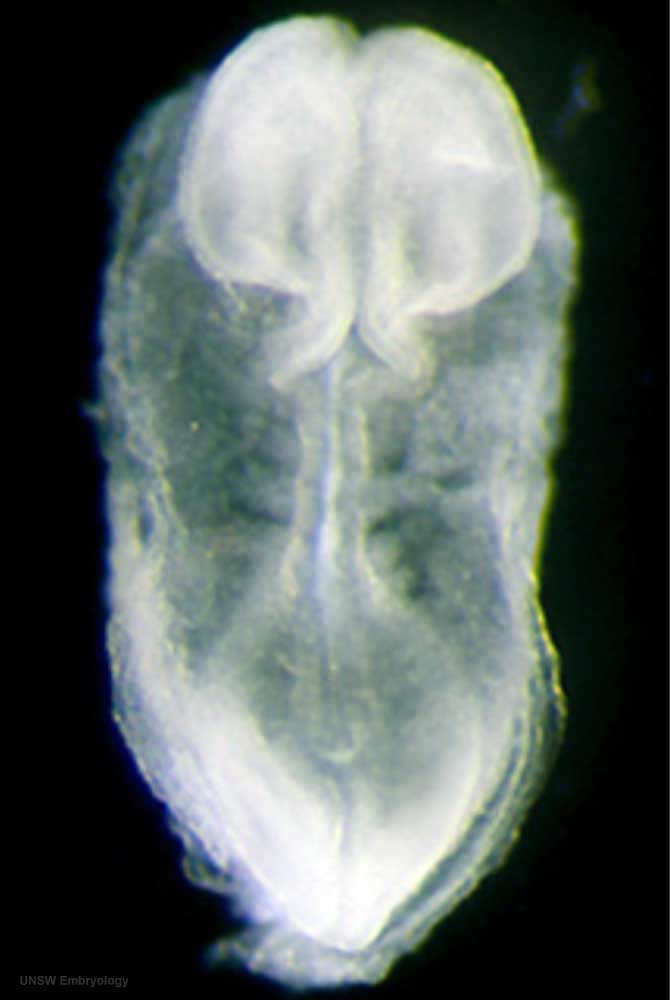 | 670 × 1,000 (27 KB) | S8600021 (talk | contribs) | == Human embryo == Stage 9 day 20, somites 3-4 Ventrolateral view (cut away from chorion and amniotic sac) showing the embryo and yolk sac. Note: # the relative size of the embryo and the associated extra-embryonic coeloms. # the shape of the early fo |
You cannot overwrite this file.
File usage
The following 5 pages use this file: