File:Stage9 bf1a.jpg
From Embryology

Size of this preview: 531 × 599 pixels. Other resolution: 709 × 800 pixels.
Original file (709 × 800 pixels, file size: 28 KB, MIME type: image/jpeg)
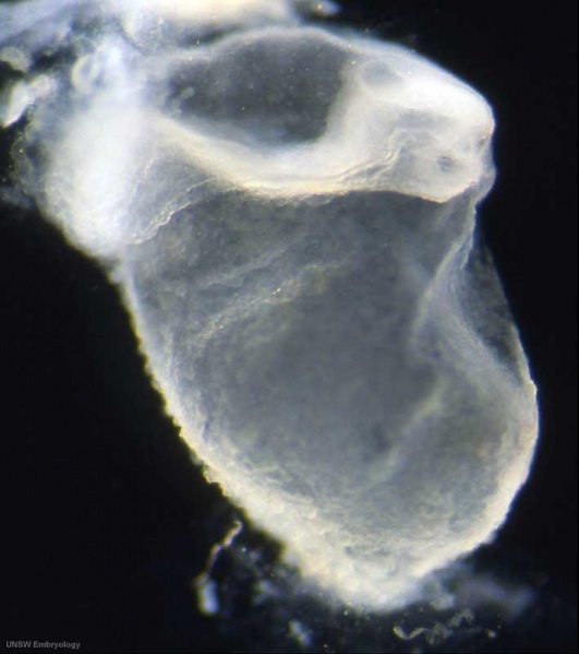
Human embryo
Stage 9 day 20, somites 3-4
Ventrolateral view (cut chorionic) showing the embryo, yolk sac and amniotic sac.
Note:
- the relative size of the embryo and the associated extra-embryonic coeloms.
- the shape of the early folded embryonic disc and rostro-caudal bendings.
- the caudal attachment of the embryo to the chorionic wall.
- the relative thicknesses of the embryo and extra-embryonic membranes.
Original file name: Stage9day20somites3-4lateralbf1-800px.jpg
Image Source: Scanning electron micrographs of the Carnegie stages of the early human embryos are reproduced with the permission of Prof Kathy Sulik, from embryos collected by Dr. Vekemans and Tania Attié-Bitach. Images are for educational purposes only and cannot be reproduced electronically or in writing without permission.
File history
Click on a date/time to view the file as it appeared at that time.
| Date/Time | Thumbnail | Dimensions | User | Comment | |
|---|---|---|---|---|---|
| current | 11:43, 22 August 2009 | | 709 × 800 (28 KB) | S8600021 (talk | contribs) | Human embryo Stage 9 day 20, somites 3-4 Ventrolateral view (cut chorionic) showing the embryo, yolk sac and amniotic sac. Note: # the relative size of the embryo and the associated extra-embryonic coeloms. # the shape of the early folded embryonic di |
You cannot overwrite this file.
File usage
The following 2 pages use this file: