File:Stage8 sem6.jpg: Difference between revisions
From Embryology
No edit summary |
|||
| (3 intermediate revisions by the same user not shown) | |||
| Line 1: | Line 1: | ||
==Human Embryo Stage 8== | ==Human Embryo Stage 8== | ||
* Carnegie stage 8, day 18 | * Carnegie stage 8, day 18 | ||
* Scanning EM dorsal view (cut amnion) showing detailed view of the prechordal region. | |||
* The mesoderm in this region will give rise to the embryonic heart tube. | |||
* Selected region from the image [[:File:Stage8 sem5.jpg|selected rostral (cranial) region]], which is in turn the rostral region of the [[:File:Stage8 sem4.jpg|entire embryonic disc]] image. | * Selected region from the image [[:File:Stage8 sem5.jpg|selected rostral (cranial) region]], which is in turn the rostral region of the [[:File:Stage8 sem4.jpg|entire embryonic disc]] image. | ||
* Note the rostro-caudal axis (top to bottom), the growing brain region of the neural plate (top) and shape of the folding embryonic disc. | * Note the rostro-caudal axis (top to bottom), the growing brain region of the neural plate (top) and shape of the folding embryonic disc. | ||
* Note the cut edge of amniotic sac with two layers (upper ectoderm, lower extra-embryonic mesoderm) | |||
:'''Links:''' [[Carnegie stage 8]] | [[:File:Stage8 sem5.jpg|selected rostral (cranial) region]] | [[:File:Stage8 sem4.jpg|entire embryonic disc]] | |||
{{Template:SEM}} | {{Template:SEM}} | ||
[[Category:Carnegie Stage]] [[Category:Carnegie Stage 8]][[Category:Heart]] | [[Category:Carnegie Stage]] [[Category:Carnegie Stage 8]][[Category:Heart]] | ||
Latest revision as of 06:41, 22 November 2011
Human Embryo Stage 8
- Carnegie stage 8, day 18
- Scanning EM dorsal view (cut amnion) showing detailed view of the prechordal region.
- The mesoderm in this region will give rise to the embryonic heart tube.
- Selected region from the image selected rostral (cranial) region, which is in turn the rostral region of the entire embryonic disc image.
- Note the rostro-caudal axis (top to bottom), the growing brain region of the neural plate (top) and shape of the folding embryonic disc.
- Note the cut edge of amniotic sac with two layers (upper ectoderm, lower extra-embryonic mesoderm)
Image Source: Scanning electron micrographs of the Carnegie stages of the early human embryos are reproduced with the permission of Prof Kathy Sulik, from embryos collected by Dr. Vekemans and Tania Attié-Bitach. Images are for educational purposes only and cannot be reproduced electronically or in writing without permission.
File history
Click on a date/time to view the file as it appeared at that time.
| Date/Time | Thumbnail | Dimensions | User | Comment | |
|---|---|---|---|---|---|
| current | 09:36, 22 August 2009 | 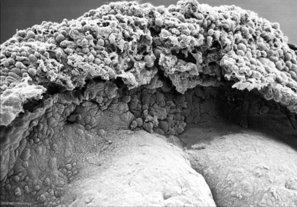 | 1,000 × 698 (87 KB) | S8600021 (talk | contribs) | Human embryo (Stage 8, day 18) Scanning EM dorsal view (cut amnion) showing detailed view of the prechordal region. Note the rostro-caudal axis (top to bottom), the growing brain region of the neural plate (top) and shape of the folding embryonic disc. |
You cannot overwrite this file.
File usage
The following 4 pages use this file: