File:Stage8 sem2.jpg: Difference between revisions
mNo edit summary |
|||
| (One intermediate revision by the same user not shown) | |||
| Line 1: | Line 1: | ||
==Scanning EM showing | ==Human Scanning EM showing the Neurenteric Canal== | ||
Human embryo ([[Carnegie stage 8|Stage 8]], day 18) showing the neurenteric canal and nodal cilia. | Human embryo ([[Carnegie stage 8|Stage 8]], day 18) showing the neurenteric canal and nodal cilia. | ||
| Line 8: | Line 8: | ||
<br><br> | |||
{{Carnegie stage 8 links}} | |||
<br><br> | |||
{{SEM}} | |||
{SEM}} | {SEM}} | ||
Latest revision as of 08:55, 23 August 2018
Human Scanning EM showing the Neurenteric Canal
Human embryo (Stage 8, day 18) showing the neurenteric canal and nodal cilia.
The neurenteric canal (archenteric canal, blastoporic canal, Braun's canal) is a transient communication that occurs briefly between the neural tube, notochordal canal, and gut endoderm.
The nodal cilia are thought to act to "stir" the overlying fluid layer at the time of gastrulation. This fluid contains secreted morphogens that are moved in one direction and therefore may have a role in establishing the initial embryo left/right axis.
Image Source: Scanning electron micrographs of the Carnegie stages of the early human embryos are reproduced with the permission of Prof Kathy Sulik, from embryos collected by Dr. Vekemans and Tania Attié-Bitach. Images are for educational purposes only and cannot be reproduced electronically or in writing without permission.
{SEM}}
Original file name:Stage8day18neuroentericcanal.jpg
File history
Click on a date/time to view the file as it appeared at that time.
| Date/Time | Thumbnail | Dimensions | User | Comment | |
|---|---|---|---|---|---|
| current | 08:58, 22 August 2009 | 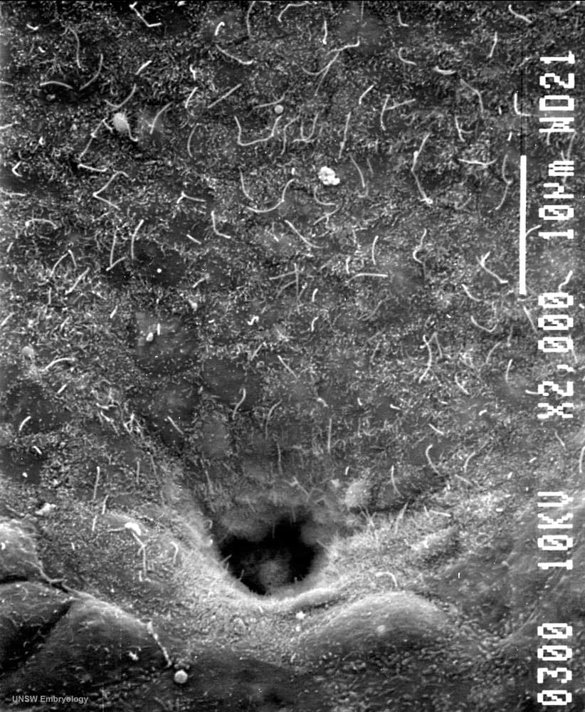 | 822 × 1,000 (142 KB) | S8600021 (talk | contribs) | Human embryo (Stage 8, day 18) Scanning EM showing detail of the neuroenteric canal. Original file name:Stage8day18neuroentericcanal.jpg {{Template:SEM}} |
You cannot overwrite this file.
File usage
The following 4 pages use this file: