File:Stage7 cloacal-oral-membranes.jpg: Difference between revisions
mNo edit summary |
mNo edit summary |
||
| Line 1: | Line 1: | ||
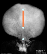
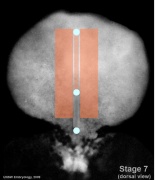
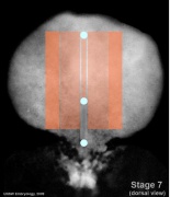
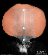
Carnegie Stages 7 showing approximate regions where buccopharyngeal and cloacal membranes would lie on the embryonic disc. | Carnegie Stages 7 showing approximate regions where buccopharyngeal and cloacal membranes would lie on the embryonic disc. | ||
{| | {| | ||
| | | | ||
* <font color=skyblue>'''top dot'''</font> - buccopharyngeal membrane | |||
* <font color=skyblue>'''top dot'''</font>- buccopharyngeal membrane | |||
* <font color=skyblue>'''middle dot'''</font> - primitive node | * <font color=skyblue>'''middle dot'''</font> - primitive node | ||
* <font color=skyblue>'''bottom dot'''</font> - cloacal membrane | * <font color=skyblue>'''bottom dot'''</font> - cloacal membrane | ||
* <font color=darkgray>'''grey line'''</font> - primitive streak | * <font color=darkgray>'''grey line'''</font> - primitive streak | ||
|- | |||
| Features: embryonic disc, primitive node, primative streak, primative groove, yolk sac | | Features: embryonic disc, primitive node, primative streak, primative groove, yolk sac | ||
Latest revision as of 13:07, 1 September 2014
Carnegie Stages 7 showing approximate regions where buccopharyngeal and cloacal membranes would lie on the embryonic disc.
|
| Features: embryonic disc, primitive node, primative streak, primative groove, yolk sac
Facts: Week 3, 15 - 17 days, 0.4 mm View 1: embryonic disc, showing the epiblast viewed from the amniotic (dorsal) side. Stage 7 Labelled | Stage 7 Molecular | Stage 7 SEM | Stage 8 Events: Gastrulation is continuing as cells migrate from the epiblast, continuing to form mesoderm. Mesoderm lies between the ectoderm and endoderm as a continuous sheet except at the buccopharyngeal and cloacal membranes. These membranes have ectoderm and endoderm only and will lie at the rostral (head) and caudal (tail) of the gastrointestinal tract. From the primitive node a tube extends under the ectoderm in the opposite direction to the primitive streak. This tube forms first the axial process then notochordal process, then finally the notochord. The notochord is a key to embryonic folding and regulation of ectoderm and mesoderm differentiation. It lies in the rostrocordal axis and the embryonic disc will fold either side ventrally, pinching off a portion of the yolk sac to form the lining of the gastrointestinal tract. |
- Embryo Stage 7 (dorsal)
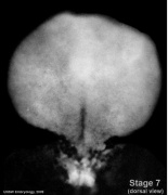
Dorsal view
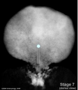
Primitive streak and node
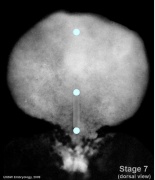
Oral and cloacal membranes
Axial mesoderm
Paraxial mesoderm
Intermediate mesoderm
Lateral plate


Reference
Image Source: UNSW Embryology
No image reuse without permission.
File history
Click on a date/time to view the file as it appeared at that time.
| Date/Time | Thumbnail | Dimensions | User | Comment | |
|---|---|---|---|---|---|
| current | 11:15, 10 August 2009 | 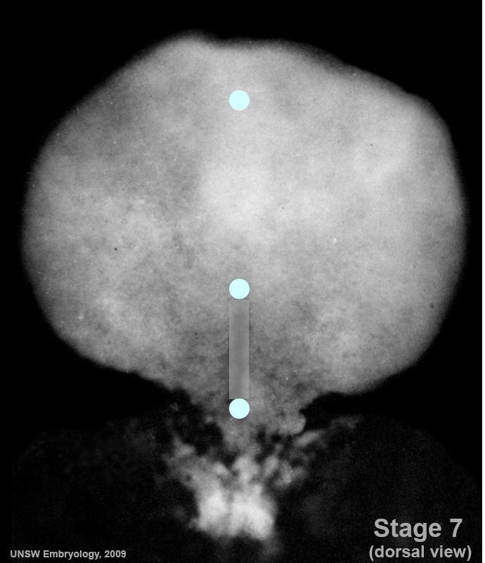 | 690 × 800 (70 KB) | MarkHill (talk | contribs) | Carnegie Stages 7 showing approximate regions where buccopharyngeal and cloacal membranes would lie on the embryonic disc. Features: embryonic disc, primitive node, primative streak, primative groove, yolk sac Facts: Week 3, 15 - 17 days, 0.4 mm View 1 |
You cannot overwrite this file.
File usage
The following 19 pages use this file:
- 2009 Lecture 5
- 2010 BGD Lecture - Development of the Embryo/Fetus 2
- 2010 Lecture 5
- BGDA Lecture - Development of the Embryo/Fetus 2
- Lecture - Gastrointestinal Development
- Lecture - Mesoderm Development
- Lecture - Week 3 Development
- Mesoderm
- Talk:2010 BGD Practical 6 - Week 3
- File:Stage7-sem2.jpg
- File:Stage7 800x700px.jpg
- File:Stage7 cloacal-oral-membranes.jpg
- File:Stage7 intermediate-mesoderm.jpg
- File:Stage7 lateral-plate.jpg
- File:Stage7 mesoderm.jpg
- File:Stage7 notochord.jpg
- File:Stage7 paraxial-mesoderm.jpg
- File:Stage7 primitive-streak-node.jpg
- Template:Stage 7 mesoderm images