File:Stage7 axial process.jpg: Difference between revisions
No edit summary |
No edit summary |
||
| Line 15: | Line 15: | ||
'''Image Source:''' UNSW Embryology, no reproduction without permission. [http://embryology.med.unsw.edu.au/wwwhuman/Stages/stage7.htm UNSW Embryology Carnegie Stage 7] | '''Image Source:''' UNSW Embryology, no reproduction without permission. [http://embryology.med.unsw.edu.au/wwwhuman/Stages/stage7.htm UNSW Embryology Carnegie Stage 7] | ||
[[Category:Human Embryo]] [[Category:Carnegie Stage]] [[Category:Carnegie Stage 7]] [[Category:Week 3]] | [[Category:Human Embryo]] [[Category:Carnegie Stage]] [[Category:Carnegie Stage 7]] [[Category:Week 3]] [[Category:Notochord]] | ||
Revision as of 15:31, 2 August 2010
Carnegie Stages 7 showing direction of axial process extension from primitive node.
Features: embryonic disc, primitive node, primative streak, primitive groove, yolk sac
Facts: Week 3, 15 - 17 days, 0.4 mm
View 1: embryonic disc, showing the epiblast viewed from the amniotic (dorsal) side.
Events: From the primitive node a tube extends under the ectoderm in the opposite direction to the primitive streak. This tube forms first the axial process, then notochordal process, and finally the notochord.
The notochord is a key to embryonic folding and regulation of ectoderm and mesoderm differentiation. It lies in the rostrocordal axis and the embryonic disc will fold either side ventrally, pinching off a portion of the yolk sac to form the lining of the gastrointestinal tract.
Original file name: Stage7n3.jpg
Image Source: UNSW Embryology, no reproduction without permission. UNSW Embryology Carnegie Stage 7
File history
Click on a date/time to view the file as it appeared at that time.
| Date/Time | Thumbnail | Dimensions | User | Comment | |
|---|---|---|---|---|---|
| current | 14:56, 3 August 2009 | 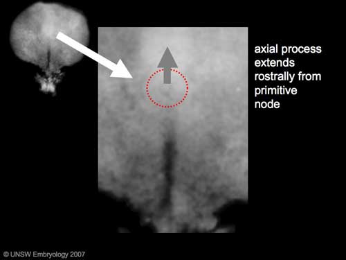 | 500 × 375 (11 KB) | MarkHill (talk | contribs) | Carnegie Stages 7 showing direction of axial process extension from primitive node. Features: embryonic disc, primitive node, primative streak, primitive groove, yolk sac Facts: Week 3, 15 - 17 days, 0.4 mm View 1: embryonic disc, showing the epiblast |
You cannot overwrite this file.
File usage
The following 9 pages use this file: