File:Stage7-bf3.jpg: Difference between revisions
| Line 6: | Line 6: | ||
Chorionic cavity (cut) showing the yolk sac (small circular structure) within the chorionic cavity. Note the relative sizes of the extraembryonic coeloms (cavities) and the attachment close to one wall of the chorionic cavity. | Chorionic cavity (cut) showing the yolk sac (small circular structure) within the chorionic cavity. Note the relative sizes of the extraembryonic coeloms (cavities) and the attachment close to one wall of the chorionic cavity. | ||
:'''Links:''' [[Carnegie stage 7]] | |||
{{Carnegie stages}} | {{Carnegie stages}} | ||
Revision as of 06:39, 18 November 2011
Human Embryo Carnegie stage 7
17 days, pre-somite, bright field image
High power image of selected region showing yolk sac and chorionic cavity from full image.
Chorionic cavity (cut) showing the yolk sac (small circular structure) within the chorionic cavity. Note the relative sizes of the extraembryonic coeloms (cavities) and the attachment close to one wall of the chorionic cavity.
- Links: Carnegie stage 7
- Carnegie Stages: 1 | 2 | 3 | 4 | 5 | 6 | 7 | 8 | 9 | 10 | 11 | 12 | 13 | 14 | 15 | 16 | 17 | 18 | 19 | 20 | 21 | 22 | 23 | About Stages | Timeline
Image Source: Scanning electron micrographs of the Carnegie stages of the early human embryos are reproduced with the permission of Prof Kathy Sulik, from embryos collected by Dr. Vekemans and Tania Attié-Bitach. Images are for educational purposes only and cannot be reproduced electronically or in writing without permission.
File history
Click on a date/time to view the file as it appeared at that time.
| Date/Time | Thumbnail | Dimensions | User | Comment | |
|---|---|---|---|---|---|
| current | 12:54, 21 August 2009 | 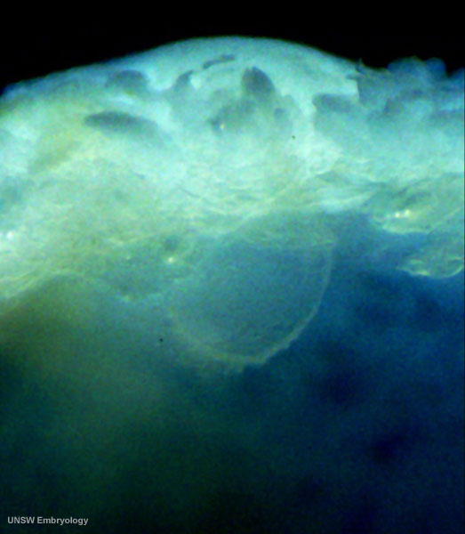 | 523 × 600 (45 KB) | MarkHill (talk | contribs) | Human Embryo Carnegie stage 7, 17 days, pre-somite, bright field image High power image of selected region from Stage7-bf2.jpg Chorionic cavity (cut) showing the yolk sac (small circular structure) within the chorionic cavity. Note |
You cannot overwrite this file.
File usage
The following 5 pages use this file: