File:Stage5 bf07.jpg
From Embryology
Size of this preview: 257 × 599 pixels. Other resolution: 304 × 708 pixels.
Original file (304 × 708 pixels, file size: 31 KB, MIME type: image/jpeg)
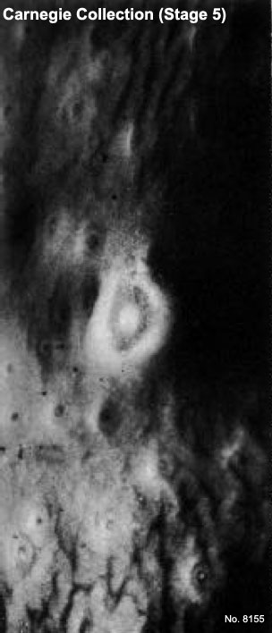
Human Embryo Carnegie Stage 5
This image shows the surface view of the site of implantation in the human uterus.
Carnegie Collection Embryo No. 8155 (stage 5a)
Facts: Week 1-2, size 0.1 - 0.2 mm
Features: implantation completed, inner cell mass, bilaminar embryo, trophoblast development
- Stage 5 Links: Embryo No.8004 - Surface view | Embryo No.8004 - near implantation site | Embryo No.8004 - Stage 5b | Embryo No.8004 - Stage 5b labeled | Embryo No.8004 - Rotated view | Embryo No.7700 - implantation site | Embryo No.7700 - Stage 5c | Implantation | Week 2
- Carnegie Stages: 1 | 2 | 3 | 4 | 5 | 6 | 7 | 8 | 9 | 10 | 11 | 12 | 13 | 14 | 15 | 16 | 17 | 18 | 19 | 20 | 21 | 22 | 23 | About Stages | Timeline
Cite this page: Hill, M.A. (2024, April 18) Embryology Stage5 bf07.jpg. Retrieved from https://embryology.med.unsw.edu.au/embryology/index.php/File:Stage5_bf07.jpg
- © Dr Mark Hill 2024, UNSW Embryology ISBN: 978 0 7334 2609 4 - UNSW CRICOS Provider Code No. 00098G
File history
Click on a date/time to view the file as it appeared at that time.
| Date/Time | Thumbnail | Dimensions | User | Comment | |
|---|---|---|---|---|---|
| current | 18:39, 23 September 2011 | | 304 × 708 (31 KB) | S8600021 (talk | contribs) | ==Human Embryo Carnegie Stage 5== Carnegie Collection Embryo No.8155 (stage 5a) Facts: Week 1-2, size 0.1 - 0.2 mm Features: implantation completed, inner cell mass, bilaminar embryo, trophoblast development {{Carnegie Embryo Stage5}} {{Template:Car |
You cannot overwrite this file.