File:Stage5 bf07.jpg
Original file (304 × 708 pixels, file size: 31 KB, MIME type: image/jpeg)
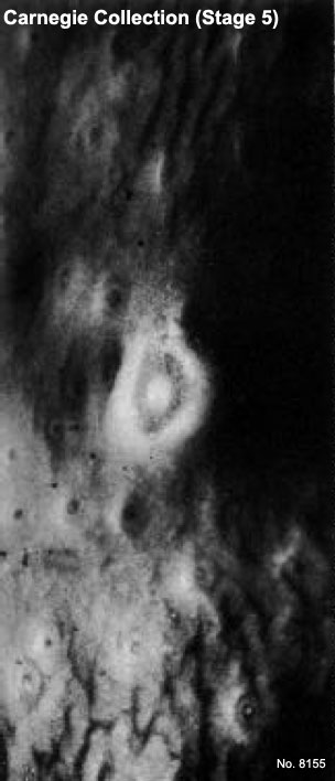
Human Embryo Carnegie Stage 5
This image shows the surface view of the site of implantation of the conceptus (pale circular region in centre of image) in the human uterus. Note the thin overlying surface epithelium and the dark ring representing the early maternal blood space.
Away from the site of implantation uterine gland opening onto the uterus surface are visible.
Carnegie Collection Embryo No. 8155 (stage 5a)
Facts: Week 1-2, size 0.1 - 0.2 mm
Features: implantation completed, inner cell mass, bilaminar embryo, trophoblast development
- Stage 5 Links: Embryo No.8004 - Surface view | Embryo No.8004 - near implantation site | Embryo No.8004 - Stage 5b | Embryo No.8004 - Stage 5b labeled | Embryo No.8004 - Rotated view | Embryo No.7700 - implantation site | Embryo No.7700 - Stage 5c | Implantation | Week 2
- Carnegie Stages: 1 | 2 | 3 | 4 | 5 | 6 | 7 | 8 | 9 | 10 | 11 | 12 | 13 | 14 | 15 | 16 | 17 | 18 | 19 | 20 | 21 | 22 | 23 | About Stages | Timeline
Cite this page: Hill, M.A. (2024, April 19) Embryology Stage5 bf07.jpg. Retrieved from https://embryology.med.unsw.edu.au/embryology/index.php/File:Stage5_bf07.jpg
- © Dr Mark Hill 2024, UNSW Embryology ISBN: 978 0 7334 2609 4 - UNSW CRICOS Provider Code No. 00098G
File history
Click on a date/time to view the file as it appeared at that time.
| Date/Time | Thumbnail | Dimensions | User | Comment | |
|---|---|---|---|---|---|
| current | 18:39, 23 September 2011 | | 304 × 708 (31 KB) | S8600021 (talk | contribs) | ==Human Embryo Carnegie Stage 5== Carnegie Collection Embryo No.8155 (stage 5a) Facts: Week 1-2, size 0.1 - 0.2 mm Features: implantation completed, inner cell mass, bilaminar embryo, trophoblast development {{Carnegie Embryo Stage5}} {{Template:Car |
You cannot overwrite this file.