File:Stage16 embryo and brain 01.jpg: Difference between revisions
No edit summary |
mNo edit summary |
||
| (5 intermediate revisions by the same user not shown) | |||
| Line 1: | Line 1: | ||
==Human Embryo Carnegie Stage16 and Neural Tube Model== | |||
The embryo and how the brain and spinal cord appears during week 6 after fertilisation (Gestational Age - GA week 8). The associated detailed description below is not part of the K12 presentation. | |||
* '''Left''' - surface view of a human embryo [[Carnegie stage 15|Week 6]]. | |||
* '''Right''' - model of neural tube surface appearance at this time and viewed also from the same direction. Note the overall shape and relative sizes of different brain regions. The neural tube is still a thin-walled tube and is mainly a fluid-filled sac. The model triangular pale region corresponds to the similar shaped dark region on the embryo. This is the very thin-walled region of the fourth (4<sup>th</sup>) ventricle. | |||
:'''Brain and Neural Tube Links:''' [[K12 Brain Awareness Week]] | [[:File:Stage15 embryo and brain 01.jpg|Week 5]] | [[:File:Stage16 embryo and brain 01.jpg|Week 6]] | [[:File:Stage22_embryo_and_brain_01.jpg|Week 8]] | [[Neural System Development|Neural Development]] | |||
'''''Gestational Age''' - is the clinical staging term of human development measured from the last menstrual period (indicating when the mother was not pregnant) not from fertilisation (about the time of ovulation of the next cycle).'' | |||
<br> | |||
{{Carnegie stage 16 links}} | |||
<br> | |||
{{Carnegie_stage_table_1}} | |||
<br> | |||
{{Footer}} | |||
[[Category:Human]] [[Category:Week 6]] [[Category:Carnegie Stage 16]] [[Category:Neural]] | |||
Latest revision as of 15:47, 24 May 2017
Human Embryo Carnegie Stage16 and Neural Tube Model
The embryo and how the brain and spinal cord appears during week 6 after fertilisation (Gestational Age - GA week 8). The associated detailed description below is not part of the K12 presentation.
- Left - surface view of a human embryo Week 6.
- Right - model of neural tube surface appearance at this time and viewed also from the same direction. Note the overall shape and relative sizes of different brain regions. The neural tube is still a thin-walled tube and is mainly a fluid-filled sac. The model triangular pale region corresponds to the similar shaped dark region on the embryo. This is the very thin-walled region of the fourth (4th) ventricle.
- Brain and Neural Tube Links: K12 Brain Awareness Week | Week 5 | Week 6 | Week 8 | Neural Development
Gestational Age - is the clinical staging term of human development measured from the last menstrual period (indicating when the mother was not pregnant) not from fertilisation (about the time of ovulation of the next cycle).
| Week: | 1 | 2 | 3 | 4 | 5 | 6 | 7 | 8 |
| Carnegie stage: | 1 2 3 4 | 5 6 | 7 8 9 | 10 11 12 13 | 14 15 | 16 17 | 18 19 | 20 21 22 23 |
Cite this page: Hill, M.A. (2024, April 18) Embryology Stage16 embryo and brain 01.jpg. Retrieved from https://embryology.med.unsw.edu.au/embryology/index.php/File:Stage16_embryo_and_brain_01.jpg
- © Dr Mark Hill 2024, UNSW Embryology ISBN: 978 0 7334 2609 4 - UNSW CRICOS Provider Code No. 00098G
File history
Click on a date/time to view the file as it appeared at that time.
| Date/Time | Thumbnail | Dimensions | User | Comment | |
|---|---|---|---|---|---|
| current | 15:55, 13 March 2012 | 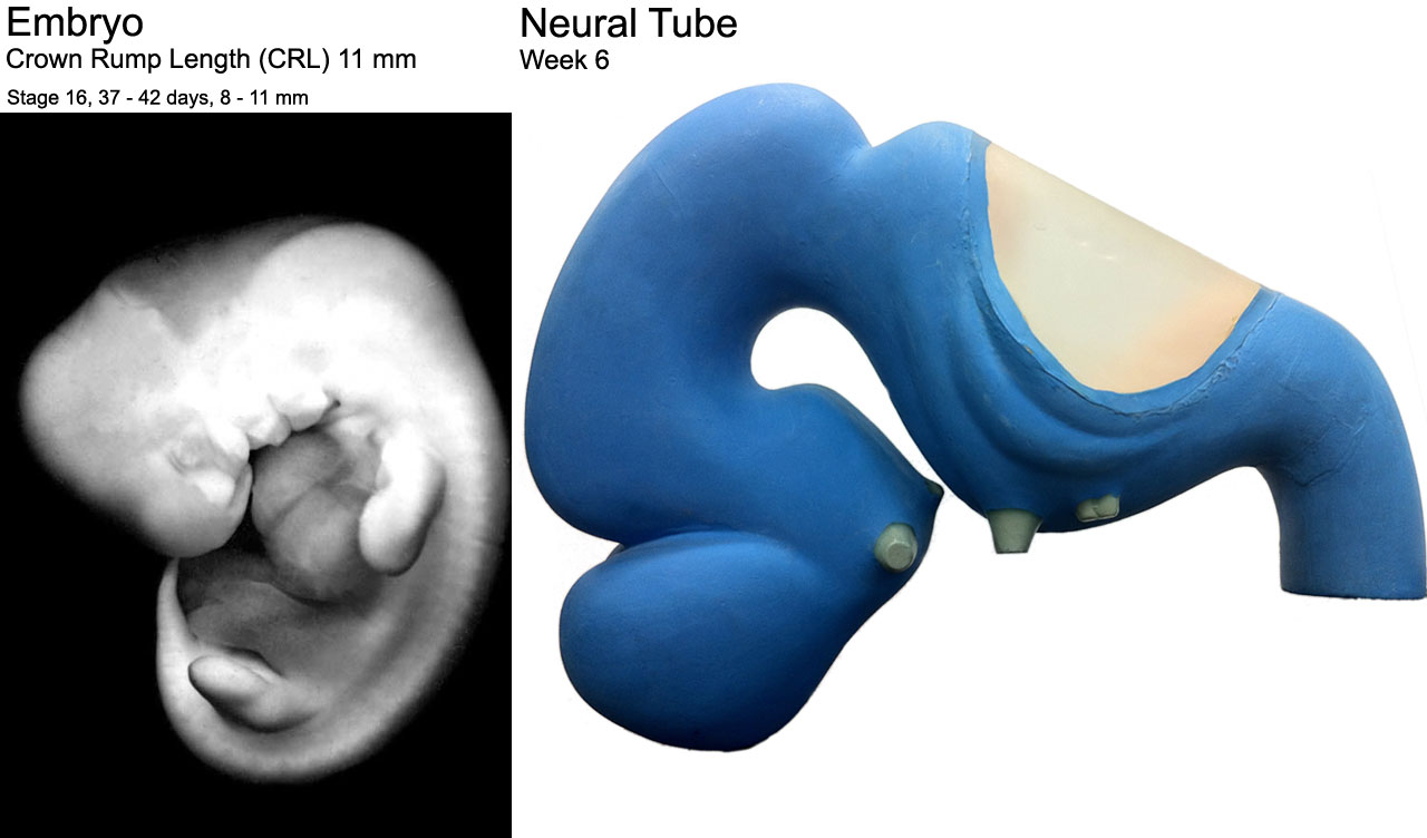 | 1,280 × 752 (91 KB) | Z8600021 (talk | contribs) |
You cannot overwrite this file.
File usage
The following 4 pages use this file: