File:Stage14compare23.jpg: Difference between revisions
From Embryology
No edit summary |
No edit summary |
||
| Line 1: | Line 1: | ||
==Human Embryo Size Comparison== | |||
'''Carnegie Stage 14 | The image compares the size of the embryo at 5 weeks (Stage 14) to that at 8 weeks (Stage 32). These stages represent approximately the middle and at the end of the embryonic period. The embryo images have been scaled to the same magnification (5 mm scale bar). | ||
:'''Links:''' [[Carnegie stage 14]] | [[Carnegie stage 23]] | [[Week 5]] | [[Week 8]] | [[Embryonic Development]] | |||
===Week 5 - Carnegie Stage 14=== | |||
* Facts: Week 5, 31 - 35 days, 5 - 7 mm | * Facts: Week 5, 31 - 35 days, 5 - 7 mm | ||
* View: Lateral view. Amniotic membrane removed. | * View: Lateral view. Amniotic membrane removed. | ||
| Line 8: | Line 14: | ||
===Week 8 - Carnegie Stage 23=== | |||
* Facts: Week 8, 56 - 60 days, 27 - 31 mm | * Facts: Week 8, 56 - 60 days, 27 - 31 mm | ||
* View: Lateral view. Amniotic membrane removed. | * View: Lateral view. Amniotic membrane removed. | ||
| Line 14: | Line 21: | ||
* Original Image Source: http://embryology.med.unsw.edu.au/wwwhuman/Stages/Images/stage14compare23.jpg | * Original Image Source: http://embryology.med.unsw.edu.au/wwwhuman/Stages/Images/stage14compare23.jpg | ||
Revision as of 11:49, 19 July 2012
Human Embryo Size Comparison
The image compares the size of the embryo at 5 weeks (Stage 14) to that at 8 weeks (Stage 32). These stages represent approximately the middle and at the end of the embryonic period. The embryo images have been scaled to the same magnification (5 mm scale bar).
- Links: Carnegie stage 14 | Carnegie stage 23 | Week 5 | Week 8 | Embryonic Development
Week 5 - Carnegie Stage 14
- Facts: Week 5, 31 - 35 days, 5 - 7 mm
- View: Lateral view. Amniotic membrane removed.
- Features: midbrain, nasal placode, lens pit, 1,2,3 pharyngeal arches, fourth ventricle of brain, 1st pharyngeal groove, heart prominence, cervical sinus, upper limb bud, mesonephric ridge, lower limb bud, umbilical cord
- Identify: midbrain region, nasal placode, lens pit, 1st, 2nd and 3rd pharyngeal arches, 1st pharyngeal groove, maxillary and mandibular components of 1st pharyngeal arch, fourth ventricle of brain, heart prominence, cervical sinus, upper limb bud, mesonephric ridge, lower limb bud, umbilical cord
Week 8 - Carnegie Stage 23
- Facts: Week 8, 56 - 60 days, 27 - 31 mm
- View: Lateral view. Amniotic membrane removed.
- Features: scalp vascular plexus, eylid, eye, nose, auricle of external ear, mouth, sholder, arm, elbow, wrist, toes separated, sole of foot, umbilical cord
- Original Image Source: http://embryology.med.unsw.edu.au/wwwhuman/Stages/Images/stage14compare23.jpg
Image Source: UNSW Embryology Carnegie Stages
© Dr Mark Hill, Image cannot be reproduced without permission.
File history
Click on a date/time to view the file as it appeared at that time.
| Date/Time | Thumbnail | Dimensions | User | Comment | |
|---|---|---|---|---|---|
| current | 20:56, 13 May 2009 | 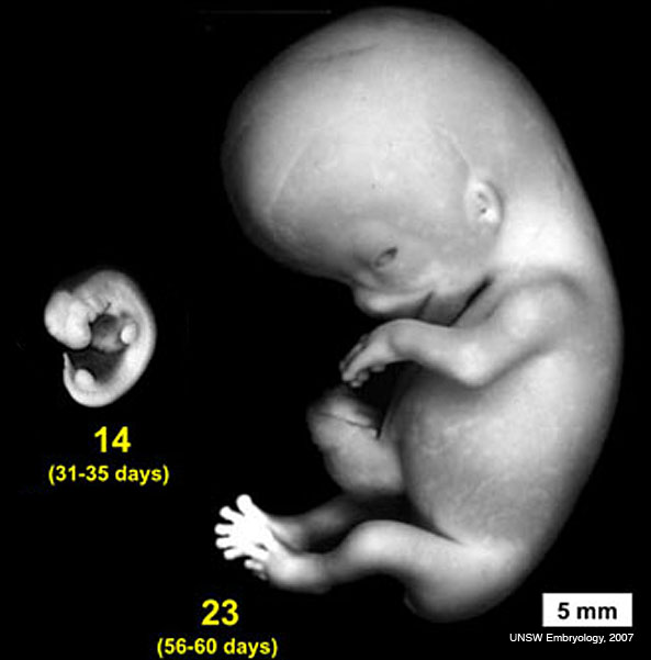 | 593 × 601 (32 KB) | MarkHill (talk | contribs) | Human Embryo comparison of embryo sizes between Stage 14 and Stage 23 http://embryology.med.unsw.edu.au/wwwhuman/Stages/Images/stage14compare23.jpg Image source: UNSW Embryology © Dr Mark Hill, Image cannot be reproduced without permission. |
You cannot overwrite this file.
File usage
The following 17 pages use this file:
- 2009 BGD-B Lecture Face and Ear
- 2009 Lecture 17
- 2010 Lecture 17
- Carnegie stage 14
- Embryonic Development
- Lecture - Sensory Development
- Sensory - Hearing and Balance Development
- Talk:Main Page
- Talk:Science
- Category:Week 1
- Category:Week 2
- Category:Week 3
- Category:Week 4
- Category:Week 5
- Category:Week 6
- Category:Week 7
- Category:Week 8