File:Stage14 sem2.jpg: Difference between revisions
No edit summary |
No edit summary |
||
| Line 1: | Line 1: | ||
==Human Embryo Carnegie stage 14== | |||
Carnegie stage 14, 32 day, 35 somite pairs | |||
View: Lateral view. Amniotic membrane removed. | |||
Scanning EM lateral view | |||
Features: midbrain, nasal placode, lens pit, 1,2,3 pharyngeal arches, fourth ventricle of brain, 1st pharyngeal groove, heart prominence, cervical sinus, upper limb bud, mesonephric ridge, lower limb bud, umbilical cord | |||
Identify: midbrain region, nasal placode, lens pit, 1st, 2nd and 3rd pharyngeal arches, 1st pharyngeal groove, maxillary and mandibular components of 1st pharyngeal arch, fourth ventricle of brain, heart prominence, cervical sinus, upper limb bud, mesonephric ridge, lower limb bud, umbilical cord | |||
[[:File:Stage14_bf1.jpg|bright Field image version]] of this image also available. | |||
'''Image version links:''' [[:File:Stage14 sem2.jpg|Large 2000px]] | [[:File:Stage14 sem2a.jpg| 1000px]] | | |||
[[:File:Stage14 sem2b.jpg|Medium 800px]] | [[:File:Stage14 sem1c.jpg|Small 400px]] | |||
Original File Name: Stage14day32somite35-lateral-sem2.jpg | |||
{{Template:SEM}} | |||
{{Template:Carnegie_stages}} | |||
{{Template:Footer}} | |||
[[Category:Week 5]] [[Category:Carnegie Stage 14]] | |||
Revision as of 16:29, 9 June 2011
Human Embryo Carnegie stage 14
Carnegie stage 14, 32 day, 35 somite pairs
View: Lateral view. Amniotic membrane removed.
Scanning EM lateral view
Features: midbrain, nasal placode, lens pit, 1,2,3 pharyngeal arches, fourth ventricle of brain, 1st pharyngeal groove, heart prominence, cervical sinus, upper limb bud, mesonephric ridge, lower limb bud, umbilical cord
Identify: midbrain region, nasal placode, lens pit, 1st, 2nd and 3rd pharyngeal arches, 1st pharyngeal groove, maxillary and mandibular components of 1st pharyngeal arch, fourth ventricle of brain, heart prominence, cervical sinus, upper limb bud, mesonephric ridge, lower limb bud, umbilical cord
bright Field image version of this image also available.
Image version links: Large 2000px | 1000px | Medium 800px | Small 400px
Original File Name: Stage14day32somite35-lateral-sem2.jpg
Image Source: Scanning electron micrographs of the Carnegie stages of the early human embryos are reproduced with the permission of Prof Kathy Sulik, from embryos collected by Dr. Vekemans and Tania Attié-Bitach. Images are for educational purposes only and cannot be reproduced electronically or in writing without permission.
- Carnegie Stages: 1 | 2 | 3 | 4 | 5 | 6 | 7 | 8 | 9 | 10 | 11 | 12 | 13 | 14 | 15 | 16 | 17 | 18 | 19 | 20 | 21 | 22 | 23 | About Stages | Timeline
Cite this page: Hill, M.A. (2024, April 24) Embryology Stage14 sem2.jpg. Retrieved from https://embryology.med.unsw.edu.au/embryology/index.php/File:Stage14_sem2.jpg
- © Dr Mark Hill 2024, UNSW Embryology ISBN: 978 0 7334 2609 4 - UNSW CRICOS Provider Code No. 00098G
File history
Click on a date/time to view the file as it appeared at that time.
| Date/Time | Thumbnail | Dimensions | User | Comment | |
|---|---|---|---|---|---|
| current | 15:06, 9 June 2011 | 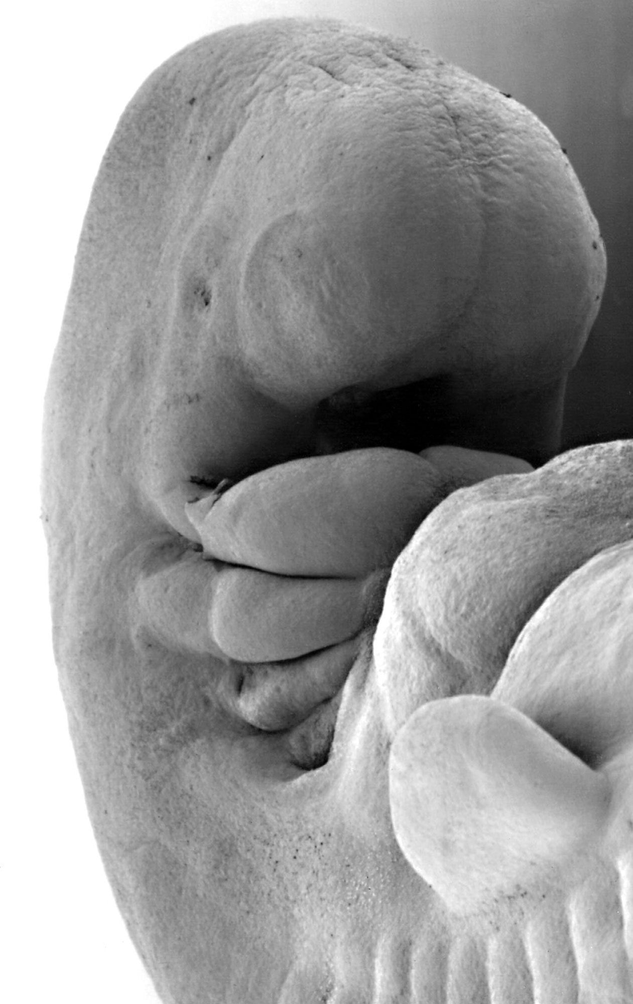 | 1,261 × 2,000 (234 KB) | S8600021 (talk | contribs) |
You cannot overwrite this file.
File usage
The following 9 pages use this file: