File:Stage14 respiratory tract.jpg: Difference between revisions
mNo edit summary |
|||
| (6 intermediate revisions by the same user not shown) | |||
| Line 1: | Line 1: | ||
==Stage14 Respiratory Tract Development== | ==Stage14 Respiratory Tract Development== | ||
This diagram is designed to show the | This diagram is designed to show the association between the foregut and the early respiratory system at about stage 13 to 14 (week 4-5) of development. | ||
Excerpts of the histology sections and their approximate level are shown in the cartoon of the embryonic respiratory/gastrointestinal tracts. | |||
The table below allows you to view each cross-section from the overview. | * initial bifurcation of foregut (oesophagus) and respiratory (trachea). | ||
* heart (ventral) and the dorsal aortas (dorsal) to the lung buds. | |||
* {{stomach}} below the lung buds. | |||
* narrow pleural canals outside the lung buds. | |||
{{Respiratory Links}} | |||
The table below allows you to view each cross-section from the above overview (section name in black text) rostrocaudally from [[:File:Stage 13 image 061.jpg|B5]] to [[:File:Stage 13 image 073.jpg|D3]]. | |||
{| | {| | ||
| Line 59: | Line 66: | ||
|} | |} | ||
{{Footer}} | |||
[[Category:Respiratory]] | [[Category:Respiratory]] | ||
Latest revision as of 21:13, 24 February 2019
Stage14 Respiratory Tract Development
This diagram is designed to show the association between the foregut and the early respiratory system at about stage 13 to 14 (week 4-5) of development.
Excerpts of the histology sections and their approximate level are shown in the cartoon of the embryonic respiratory/gastrointestinal tracts.
- initial bifurcation of foregut (oesophagus) and respiratory (trachea).
- heart (ventral) and the dorsal aortas (dorsal) to the lung buds.
- stomach below the lung buds.
- narrow pleural canals outside the lung buds.
The table below allows you to view each cross-section from the above overview (section name in black text) rostrocaudally from B5 to D3.

|

|

|

|

|

|

|
| B1L | B2L | B3L | B4L | B5L | B6L | B7L |

|

|

|

|

|

|

|
| C1L | C2L | C3L | C4L | C5L | C6L | C7L |

|

|

|

|

|

|

|
| D1L | D2L | D3L | D4L | D5L | D6L | D7L |
Cite this page: Hill, M.A. (2024, April 18) Embryology Stage14 respiratory tract.jpg. Retrieved from https://embryology.med.unsw.edu.au/embryology/index.php/File:Stage14_respiratory_tract.jpg
- © Dr Mark Hill 2024, UNSW Embryology ISBN: 978 0 7334 2609 4 - UNSW CRICOS Provider Code No. 00098G
File history
Click on a date/time to view the file as it appeared at that time.
| Date/Time | Thumbnail | Dimensions | User | Comment | |
|---|---|---|---|---|---|
| current | 14:04, 24 August 2009 | 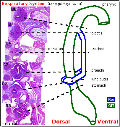 | 406 × 431 (76 KB) | MarkHill (talk | contribs) | Stage14 respiratory tract Original file name: lung2.gif Image source: UNSW Embryology http://embryology.med.unsw.edu.au/Notes/respire.htm |
You cannot overwrite this file.
File usage
The following 17 pages use this file:
- 2009 Lab 5
- 2009 Lecture 10
- 2010 BGD Lecture - Development of the Embryo/Fetus 2
- 2010 BGD Practical 6 - Week 5
- 2010 Lab 5
- 2010 Lecture 10
- 2011 Lab 5 - Early Embryo
- ANAT2341 Lab 5 - Early Embryo
- BGDA Lecture - Development of the Embryo/Fetus 2
- Draft 2016
- Human System Development
- Lecture - Respiratory Development
- R
- Respiratory System - Carnegie Stage 13
- Respiratory System - Upper Respiratory Tract
- Respiratory System Development
- SH Lecture - Respiratory System Development