File:Stage13 sem2.jpg: Difference between revisions
No edit summary |
mNo edit summary |
||
| Line 8: | Line 8: | ||
* pharyngeal arches | * pharyngeal arches | ||
* heart | * heart | ||
[[:File:Stage13_bf1.jpg|Bright Field image version]] of this image also available. | [[:File:Stage13_bf1.jpg|Bright Field image version]] of this image also available. | ||
| Line 23: | Line 19: | ||
{{ | {{SEM}} | ||
{{ | {{Carnegie_stages}} | ||
{{Footer}} | |||
[[Category:Week 5]] [[Category:Head]] [[Category:Face]] | [[Category:Week 5]] [[Category:Head]] [[Category:Face]] | ||
Revision as of 17:18, 24 July 2017
Human Embryo Carnegie stage 13
Carnegie stage 13, 28 day, 30 somite pairs
Scanning EM lateral view of head region showing detail of pharyngeal arches from full embryo SEM.
- yolk sac and amnion removed
- pharyngeal arches
- heart
Bright Field image version of this image also available.
Image version links: Large 1000px | 800px |
Medium 600px | Small 400px
Related Images: Full Embryo SEM | Bright Field image version
Image Source: Scanning electron micrographs of the Carnegie stages of the early human embryos are reproduced with the permission of Prof Kathy Sulik, from embryos collected by Dr. Vekemans and Tania Attié-Bitach. Images are for educational purposes only and cannot be reproduced electronically or in writing without permission.
- Carnegie Stages: 1 | 2 | 3 | 4 | 5 | 6 | 7 | 8 | 9 | 10 | 11 | 12 | 13 | 14 | 15 | 16 | 17 | 18 | 19 | 20 | 21 | 22 | 23 | About Stages | Timeline
Cite this page: Hill, M.A. (2024, April 24) Embryology Stage13 sem2.jpg. Retrieved from https://embryology.med.unsw.edu.au/embryology/index.php/File:Stage13_sem2.jpg
- © Dr Mark Hill 2024, UNSW Embryology ISBN: 978 0 7334 2609 4 - UNSW CRICOS Provider Code No. 00098G
File history
Click on a date/time to view the file as it appeared at that time.
| Date/Time | Thumbnail | Dimensions | User | Comment | |
|---|---|---|---|---|---|
| current | 10:22, 4 September 2009 | 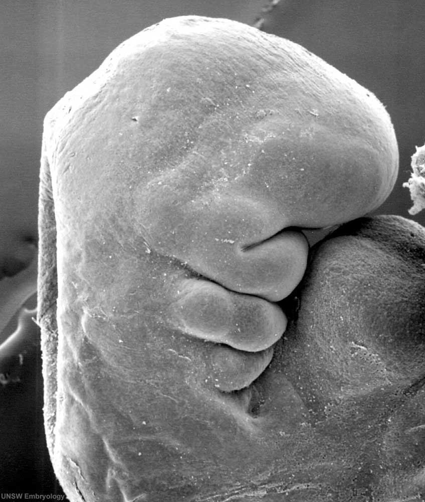 | 848 × 1,000 (84 KB) | S8600021 (talk | contribs) | '''Human Embryo''' Carnegie stage 13, 28 day, 30 somite pairs Scanning EM lateral view * yolk sac and amnion removed * pharyngeal arches * heart * limb buds Bright Field image version of this image also available. Original |
You cannot overwrite this file.
File usage
The following 11 pages use this file:
- AACP Meeting 2013 - Face Embryology
- Abnormal Development - Thalidomide
- BGD Lecture - Face and Ear Development
- Carnegie stage 13
- Hearing - Inner Ear Development
- Human Embryo - Scanning electron microscopy
- Human Embryo SEM
- K12 Thalidomide
- Lecture - Head Development
- Talk:Carnegie stage 13
- Template:Carnegie stage 11-14 image table