File:Stage13 sem1c.jpg: Difference between revisions
mNo edit summary |
|||
| Line 19: | Line 19: | ||
{{ | {{Stage13SEM}} | ||
| Line 25: | Line 25: | ||
{{ | {{SEM}} | ||
{{ | {{Carnegie_stages}} | ||
{{ | {{Footer}} | ||
[[Category:Head]] [[Category:Limb]] | [[Category:Head]] [[Category:Limb]] | ||
[[Category:Carnegie Stage 13]] [[Category:Week 4]] | [[Category:Carnegie Stage 13]] [[Category:Week 4]] | ||
Revision as of 08:31, 9 February 2016
Human Embryo Carnegie stage 13
Carnegie stage 13, 28 day, 30 somite pairs
Scanning EM lateral view
- yolk sac and amnion removed
- pharyngeal arches
- heart
- limb buds
Bright Field image version of this image also available.
Original File Name: Stage13day28somite30-lateral-arches-sem1.jpg
Image version links
Large 1000px | 800px | Medium 600px | Small 400px
Stage 13 SEM Images: embryo lateral view | head lateral view | pharyngeal arches
Related Images: Bright Field image version
Image Source: Scanning electron micrographs of the Carnegie stages of the early human embryos are reproduced with the permission of Prof Kathy Sulik, from embryos collected by Dr. Vekemans and Tania Attié-Bitach. Images are for educational purposes only and cannot be reproduced electronically or in writing without permission.
- Carnegie Stages: 1 | 2 | 3 | 4 | 5 | 6 | 7 | 8 | 9 | 10 | 11 | 12 | 13 | 14 | 15 | 16 | 17 | 18 | 19 | 20 | 21 | 22 | 23 | About Stages | Timeline
Cite this page: Hill, M.A. (2024, April 18) Embryology Stage13 sem1c.jpg. Retrieved from https://embryology.med.unsw.edu.au/embryology/index.php/File:Stage13_sem1c.jpg
- © Dr Mark Hill 2024, UNSW Embryology ISBN: 978 0 7334 2609 4 - UNSW CRICOS Provider Code No. 00098G
File history
Click on a date/time to view the file as it appeared at that time.
| Date/Time | Thumbnail | Dimensions | User | Comment | |
|---|---|---|---|---|---|
| current | 21:23, 3 September 2009 | 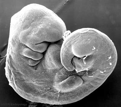 | 400 × 355 (18 KB) | S8600021 (talk | contribs) |
You cannot overwrite this file.
File usage
The following 37 pages use this file:
- 2009 Lecture 14
- 2010 BGD Lecture - Development of the Embryo/Fetus 2
- 2010 BGD Practical 6 - Week 8
- 2010 BGD Tutorial - Applied Embryology and Teratology
- 2010 Lab 6
- 2010 Lecture 14
- 2010 Lecture 6
- 2011 Group Project 11
- 2011 Lab 10 - Early Embryo
- 2011 Lab 6 - Early Embryo
- ANAT2341 Lab 10 - Early Embryo
- ANAT2341 Lab 6 - Early Embryo
- Abnormal Development - Environmental
- Abnormal Development - Illegal Drugs
- Abnormal Development - Teratogens
- BGDA Lecture - Development of the Embryo/Fetus 2
- BGDA Practical 3 - Week 3 Summary
- BGDA Practical 7 - Week 8
- BGDB Face and Ear - Early Embryo
- BGD Tutorial - Applied Embryology and Teratology
- Embryonic Development
- Fetal ECHO Meeting 2012
- Foundations Practical - Critical Periods
- Human Abnormal Development
- Human Embryo SEM
- Lecture - Ectoderm Development
- Lecture - Fetal Development
- Lecture - Limb Development
- RPAH Cardiac Embryology 2014
- Timeline human development
- Talk:Timeline human development
- File:Human-critical periods of development.jpg
- File talk:Human-critical periods.jpg
- Template:Critical Periods table
- Template:First Trimester Timeline
- Template:First Trimester Timeline collapsable table
- Template talk:First Trimester Timeline