File:Stage12 sem6.jpg: Difference between revisions
From Embryology
(== Human Embryo Carnegie Stage 12== Carnegie Stage 12 Facts: Week 4, 26 days, 5 mm, Somite Number 21 View: Lateral view, day 26, 21 somites, amniotic membrane removed Features: day 26, 27 somites, forebrain, site of lens placode, site of otic placode ,) |
mNo edit summary |
||
| Line 1: | Line 1: | ||
== Human Embryo Carnegie Stage 12== | == Human Embryo Carnegie Stage 12== | ||
Carnegie Stage 12 | * [[Carnegie Stage 12]] [[Week 4]], 26 days, 5 mm, Somite Number 21. | ||
* View: Lateral view, day 26, 21 somites, amniotic membrane removed | |||
* Features: day 26, 27 somites, forebrain, site of lens placode, site of otic placode , stomodeum, 1st pharyngeal arch, 2nd pharyngeal arch, 3rdpharyngeal arch, heart prominence, somite | |||
* Identify: forebrain, site of lens placode, site of otic placode, stomodeum, 1st pharyngeal arch, 2nd pharyngeal arch, 3rd pharyngeal arch, heart prominence, somite | |||
* [[:File:Stage12 bf2.jpg|Bright field image version]] of this image also available. | |||
{{Stage 12 SEM images}} | |||
{{SEM}} | |||
{{Carnegie_stages}} | |||
Image version links: [[:File:Stage12 sem6.jpg|Large 1600px]] | [[:File:Stage12 sem6a.jpg| 1000px]] | [[:File:Stage12 sem6b.jpg|Medium 800px]] | [[:File:Stage12 sem6c.jpg|Small 600px]] | |||
[[:File:Stage12 | |||
Original File Name: Stage12day26somite21 ventrolateral sem6.jpg | Original File Name: Stage12day26somite21 ventrolateral sem6.jpg | ||
{{Footer}} | |||
{{ | |||
[[Category:Carnegie Stage 12]] [[Category:Carnegie Stage]] [[Category:Week 4]] | [[Category:Carnegie Stage 12]] [[Category:Carnegie Stage]] [[Category:Week 4]] | ||
[[Category:Neural]] [[Category:Heart]] | [[Category:Neural]] [[Category:Heart]] | ||
Revision as of 22:42, 4 September 2014
Human Embryo Carnegie Stage 12
- Carnegie Stage 12 Week 4, 26 days, 5 mm, Somite Number 21.
- View: Lateral view, day 26, 21 somites, amniotic membrane removed
- Features: day 26, 27 somites, forebrain, site of lens placode, site of otic placode , stomodeum, 1st pharyngeal arch, 2nd pharyngeal arch, 3rdpharyngeal arch, heart prominence, somite
- Identify: forebrain, site of lens placode, site of otic placode, stomodeum, 1st pharyngeal arch, 2nd pharyngeal arch, 3rd pharyngeal arch, heart prominence, somite
- Bright field image version of this image also available.
- Stage 12 SEM Images: Bright Field 1 | Bright Field 3 | Bright Field 3 | SEM1 | SEM2 | SEM3 | SEM4 dorsolateral head and arches | SEM5 lateral head and arches | SEM6 ventrolateral head and arches | SEM7 lateral | SEM8 ventrolateral | SEM9 cloacal membrane | SEM9 labeled | Carnegie stage 12
Image Source: Scanning electron micrographs of the Carnegie stages of the early human embryos are reproduced with the permission of Prof Kathy Sulik, from embryos collected by Dr. Vekemans and Tania Attié-Bitach. Images are for educational purposes only and cannot be reproduced electronically or in writing without permission.
- Carnegie Stages: 1 | 2 | 3 | 4 | 5 | 6 | 7 | 8 | 9 | 10 | 11 | 12 | 13 | 14 | 15 | 16 | 17 | 18 | 19 | 20 | 21 | 22 | 23 | About Stages | Timeline
Image version links: Large 1600px | 1000px | Medium 800px | Small 600px
Original File Name: Stage12day26somite21 ventrolateral sem6.jpg
Cite this page: Hill, M.A. (2024, April 24) Embryology Stage12 sem6.jpg. Retrieved from https://embryology.med.unsw.edu.au/embryology/index.php/File:Stage12_sem6.jpg
- © Dr Mark Hill 2024, UNSW Embryology ISBN: 978 0 7334 2609 4 - UNSW CRICOS Provider Code No. 00098G
File history
Click on a date/time to view the file as it appeared at that time.
| Date/Time | Thumbnail | Dimensions | User | Comment | |
|---|---|---|---|---|---|
| current | 15:20, 14 May 2011 | 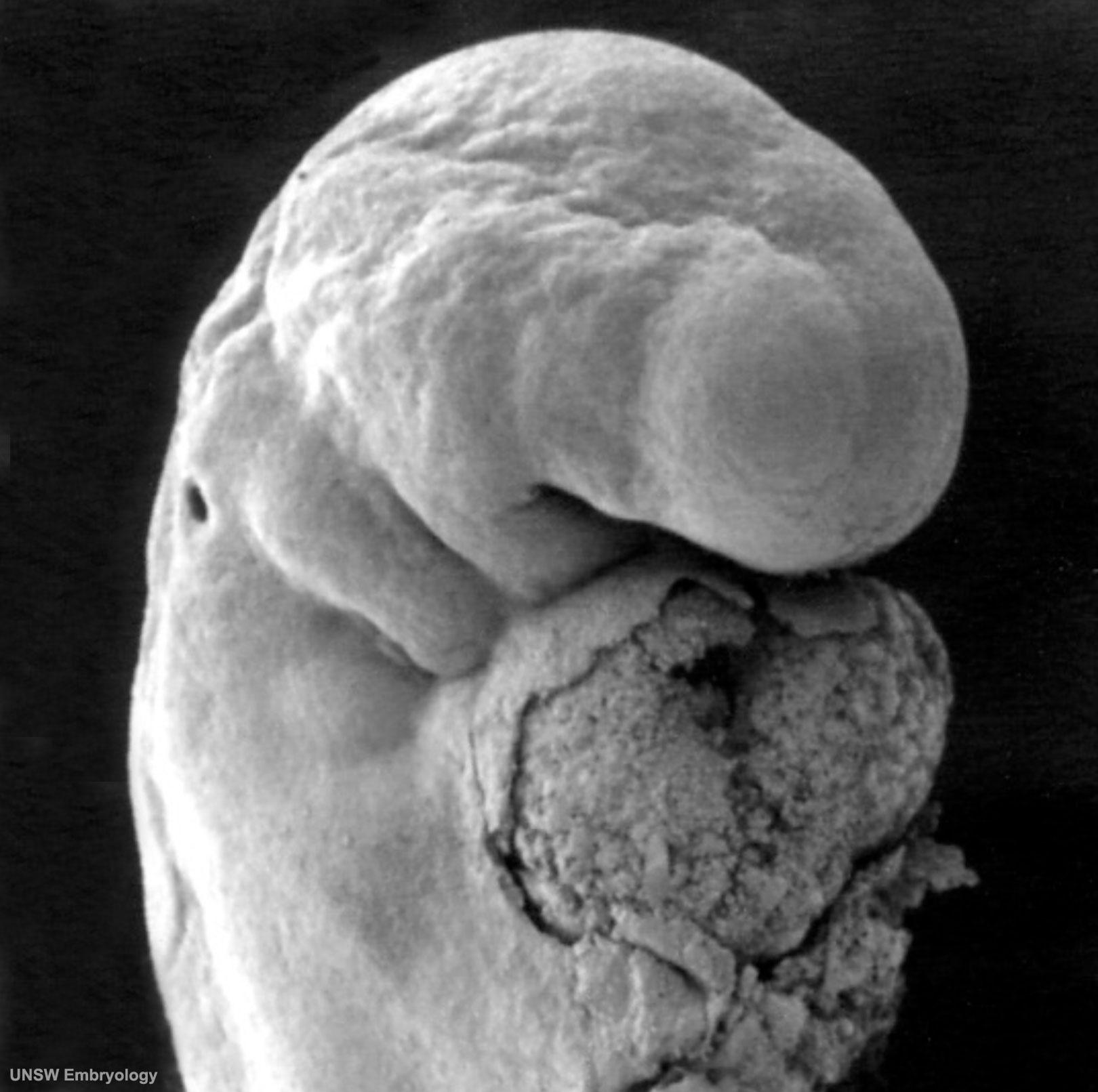 | 1,620 × 1,612 (190 KB) | S8600021 (talk | contribs) | == Human Embryo Carnegie Stage 12== Carnegie Stage 12 Facts: Week 4, 26 days, 5 mm, Somite Number 21 View: Lateral view, day 26, 21 somites, amniotic membrane removed Features: day 26, 27 somites, forebrain, site of lens placode, site of otic placode , |
You cannot overwrite this file.