File:Stage12 SEM3.jpg: Difference between revisions
No edit summary |
No edit summary |
||
| Line 1: | Line 1: | ||
Original file name: Stage12semneuropore.jpg | |||
'''Image Source:''' Prof Kathy Sulik scanning electron micrographs of the Carnegie stages of the early human embryo. UNSW Embryology, no reproduction without permission. [http://embryology.med.unsw.edu.au/wwwhuman/Stages/Stagesem.htm Carnegie Stages - Scanning Electron Micrography] | [http://embryology.med.unsw.edu.au/wwwhuman/Stages/stage12SEM.htm Embryology page Created: 2007] | |||
===Carnegie Stages Link=== | |||
[[Carnegie_stage_1|1]] | [[Carnegie_stage_3|3]] | [[Carnegie_stage_7|7]] | [[Carnegie_stage_8|8]] | [[Carnegie_stage_9|9]] | [[Carnegie_stage_10|10]] | [[Carnegie_stage_11|11]] | [[Carnegie_stage_12|12]] | [[Carnegie_stage_13|13]] | [[Carnegie_stage_14|14]] | [[Carnegie_stage_15|15]] | [[Carnegie_stage_16|16]] | [[Carnegie_stage_17|17]] | [[Carnegie_stage_18|18]] | [[Carnegie_stage_19|19]] | [[Carnegie_stage_20|20]] | [[Carnegie_stage_21|21]] | [[Carnegie_stage_22|22]] | [[Carnegie_stage_23|23]] | |||
== About Carnegie Stages == | |||
Carnegie stages are named after the famous US Institute which began collecting and classifying embryos in the early 1900's. Stages are based on the external and/or internal morphological development of the embryo, and are not directly dependent on either age or size. The human embryonic period proper is divided into 23 Carnegie stages. Carnegie stages are based on the external and/or internal morphological development of the embryo, and are not directly dependent on either age or size. Criteria beyond morphological features include age in days, number of somites present, and embryonic length. | |||
The Kyoto Collection images are reproduced with the permission of '''Prof. Kohei Shiota'''. Scanning electron micrographs of the Carnegie stages of the early human embryos are reproduced with the permission of '''Prof Kathy Sulik'''. Images are for educational tutorial/revision purposes and cannot be reproduced electronically or in writing without permission. | |||
== UNSW Embryology Links == | |||
* [http://embryology.med.unsw.edu.au/wwwhuman/Stages/stage12.htm Embryology Carnegie stage 12 Created: 19.03.1999] | |||
* [http://embryology.med.unsw.edu.au/wwwhuman/Stages/carnegie.htm About the Carnegie Institute] | |||
===Glossary Links=== | |||
[[A|A]] | [[B|B]] | [[C|C]] | [[D|D]] | [[E|E]] | [[F|F]] | [[G|G]] | [[H|H]] | [[I|I]] | [[J|J]] | [[K|K]] | [[L|L]] | [[M|M]] | [[N|N]] | [[O|O]] | [[P|P]] | [[Q|Q]] | [[R|R]] | [[S|S]] | [[T|T]] | [[U|U]] | [[V|V]] | [[W|W]] | [[X|X]] | [[Y|Y]] | [[Z|Z]] | |||
''' | :Dr Mark Hill 2009, '''''UNSW Embryology''''' ISBN: 978 0 7334 2609 4 - UNSW CRICOS Provider Code No. 00098G | ||
[[Category:Human Embryo]] [[Category:Carnegie Stage]] [[Category:Week 3]] [[Category:Scanning EM]] | [[Category:Human Embryo]] [[Category:Carnegie Stage]] [[Category:Week 4]] [[Category:Ectoderm]] [[Category:Neural]] [[Category:Week 3]] [[Category:Scanning EM]] | ||
Revision as of 11:39, 14 August 2009
Original file name: Stage12semneuropore.jpg
Image Source: Prof Kathy Sulik scanning electron micrographs of the Carnegie stages of the early human embryo. UNSW Embryology, no reproduction without permission. Carnegie Stages - Scanning Electron Micrography | Embryology page Created: 2007
Carnegie Stages Link
1 | 3 | 7 | 8 | 9 | 10 | 11 | 12 | 13 | 14 | 15 | 16 | 17 | 18 | 19 | 20 | 21 | 22 | 23
About Carnegie Stages
Carnegie stages are named after the famous US Institute which began collecting and classifying embryos in the early 1900's. Stages are based on the external and/or internal morphological development of the embryo, and are not directly dependent on either age or size. The human embryonic period proper is divided into 23 Carnegie stages. Carnegie stages are based on the external and/or internal morphological development of the embryo, and are not directly dependent on either age or size. Criteria beyond morphological features include age in days, number of somites present, and embryonic length.
The Kyoto Collection images are reproduced with the permission of Prof. Kohei Shiota. Scanning electron micrographs of the Carnegie stages of the early human embryos are reproduced with the permission of Prof Kathy Sulik. Images are for educational tutorial/revision purposes and cannot be reproduced electronically or in writing without permission.
UNSW Embryology Links
Glossary Links
A | B | C | D | E | F | G | H | I | J | K | L | M | N | O | P | Q | R | S | T | U | V | W | X | Y | Z
- Dr Mark Hill 2009, UNSW Embryology ISBN: 978 0 7334 2609 4 - UNSW CRICOS Provider Code No. 00098G
File history
Click on a date/time to view the file as it appeared at that time.
| Date/Time | Thumbnail | Dimensions | User | Comment | |
|---|---|---|---|---|---|
| current | 15:43, 10 August 2009 | 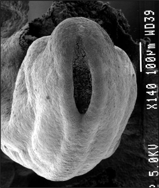 | 507 × 600 (68 KB) | MarkHill (talk | contribs) | Original file name: Stage12semneuropore.jpg |
You cannot overwrite this file.
File usage
The following 22 pages use this file:
- 2009 Lecture 6
- 2010 BGD Lecture - Development of the Embryo/Fetus 1
- 2010 BGD Lecture - Development of the Embryo/Fetus 2
- 2010 BGD Practical 6 - Week 4
- 2010 Lab 3
- 2010 Lecture 6
- 2011 Lab 3 - Week 4
- ANAT2341 Lab 3 - Week 4
- Abnormal Development - Thalidomide
- BGDA Lecture - Development of the Embryo/Fetus 1
- BGDA Lecture - Development of the Embryo/Fetus 2
- BGDA Lecture - Development of the Nervous System
- BGDA Practical 7 - Week 4
- C
- Carnegie stage 12
- Ectoderm
- Human Embryo SEM
- K12 Thalidomide
- Neural System Development
- Talk:2011 Lab 3
- Talk:BGDA Lecture - Development of the Nervous System
- Template:Carnegie stage 11-14 image table