File:Stage11 sem6.jpg: Difference between revisions
No edit summary |
|||
| Line 1: | Line 1: | ||
==Human Embryo Carnegie stage 11== | ==Human Embryo Carnegie stage 11== | ||
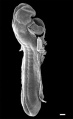
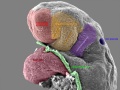
This scanning EM of the stage 11 human embryo is the same image as [[:File:Stage11 sem5.jpg|Stage11 sem5]] with the addition of | This scanning EM of the stage 11 human embryo is the same image as [[:File:Stage11 sem5.jpg|Stage11 sem5]] with the addition of direction of view. | ||
There are some alternative terminologies: | |||
* '''ventral''' and '''anterior''' both mean toward the front of the body. | |||
* '''dorsal''' and '''posterior''' mean the back of the body. | |||
* '''rostral''', '''cranial''', and '''cephalic''' and '''superior''' all mean toward the head or the upper part of a structure | |||
* proximal means closer to the trunk | |||
* distal is away from the trunk | |||
* medial describes a structure toward the midline of the body | |||
* lateral describes a structure away from the midline of the body | |||
Carnegie stage 11 24 days, 13 somite pairs | Carnegie stage 11 24 days, 13 somite pairs | ||
Revision as of 14:03, 5 May 2011
Human Embryo Carnegie stage 11
This scanning EM of the stage 11 human embryo is the same image as Stage11 sem5 with the addition of direction of view.
There are some alternative terminologies:
- ventral and anterior both mean toward the front of the body.
- dorsal and posterior mean the back of the body.
- rostral, cranial, and cephalic and superior all mean toward the head or the upper part of a structure
- proximal means closer to the trunk
- distal is away from the trunk
- medial describes a structure toward the midline of the body
- lateral describes a structure away from the midline of the body
Carnegie stage 11 24 days, 13 somite pairs
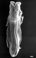
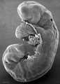
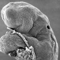
Facts: Week 4, 23 - 26 days, 2.5 - 4.5 mm, Somite Number 13 - 20
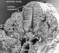
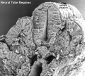
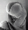
View: This is a scanning EM of the embryo dorsal view showing the neural tube closing with open neuropores and the paired somites visible through the thin ectoderm.
Features: surface ectoderm, neural tube, cranial (anterior) neuropore, caudal (posterior) neuropore, somites, heart, cut edge of amnion
Stage11_sem6.jpg
Original file name: Stage11day24somite13-dorsal-sem5-1000px.jpg
- Stage 11 SEM Images: dorsolateral whole embryo | dorsal embryo | lateral embryo | lateral head | lateral head with overlay | embryo cross-section | ventrolateral head | ventrolateral head with overlay | ventral head | buccopharyngeal membrane | neural crest | posterior neuropore | anterior neuropore | Carnegie stage 11
- Human Embryo (stage 11)
dorsolateral whole embryo
dorsal embryo
lateral embryo
lateral head
lateral head with overlay

embryo cross-section
embryo cross-section label
neural cross-section label

ventrolateral head
ventrolateral head with overlay

ventral head

buccopharyngeal membrane
neural crest
posterior neuropore
anterior neuropore


Image Source: Scanning electron micrographs of the Carnegie stages of the early human embryos are reproduced with the permission of Prof Kathy Sulik, from embryos collected by Dr. Vekemans and Tania Attié-Bitach. Images are for educational purposes only and cannot be reproduced electronically or in writing without permission.
File history
Click on a date/time to view the file as it appeared at that time.
| Date/Time | Thumbnail | Dimensions | User | Comment | |
|---|---|---|---|---|---|
| current | 14:13, 5 May 2011 | 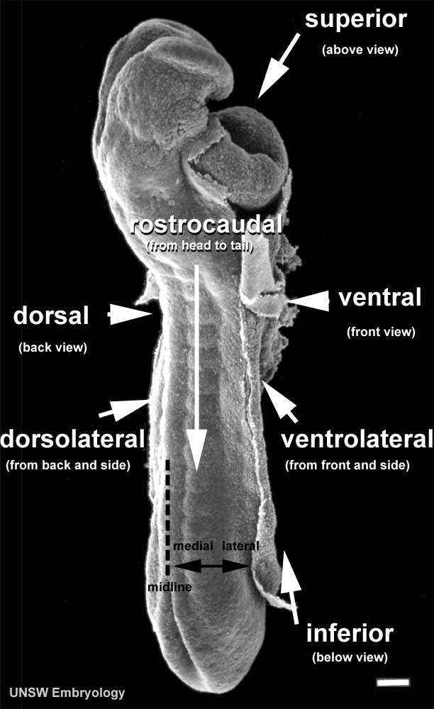 | 612 × 1,000 (57 KB) | S8600021 (talk | contribs) | |
| 13:49, 5 May 2011 | 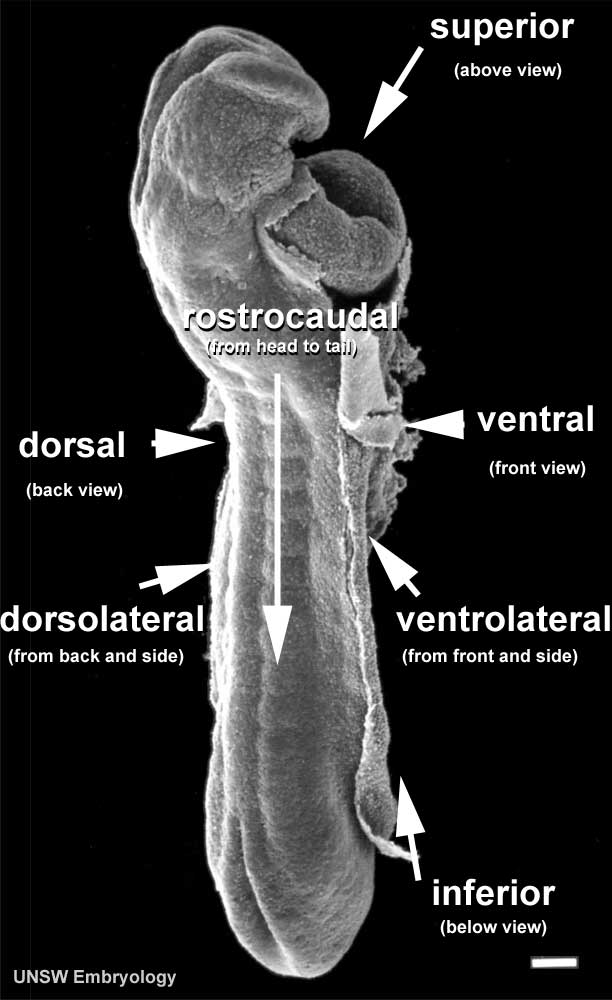 | 612 × 1,000 (57 KB) | S8600021 (talk | contribs) | ==Human Embryo Carnegie stage 11== This scanning EM of the stage 11 human embryo is the same image as Stage11 sem5 with the addition of Carnegie stage 11 24 days, 13 somite pairs Facts: Week 4, 23 - 26 days, 2.5 - 4.5 mm, So |
You cannot overwrite this file.
File usage
The following 21 pages use this file:
- 2009 Lecture 6
- 2010 BGD Lecture - Development of the Embryo/Fetus 1
- 2011 Lab 3 - Week 4
- ANAT2341 Lab 3 - Week 4
- Abnormal Development - Thalidomide
- BGDA Lecture - Development of the Embryo/Fetus 1
- BGDA Lecture - Development of the Embryo/Fetus 2
- BGDA Lecture - Development of the Nervous System
- BGDA Practical - Implantation to 8 Weeks
- BGDA Practical 7 - Week 4
- Carnegie stage 11
- Developmental Mechanism - Axes Formation
- Developmental Mechanism - Dorso-Ventral Axis
- Developmental Mechanism - Left-Right Axis
- Developmental Mechanism - Rostro-Caudal axis Axis
- Human Embryo - Scanning electron microscopy
- Human Embryo SEM
- K12 Thalidomide
- Talk:2011 Lab 3
- Talk:Carnegie stage 11
- Template:Carnegie stage 11-14 image table