File:Stage11 sem3c.jpg: Difference between revisions
No edit summary |
mNo edit summary |
||
| Line 1: | Line 1: | ||
==Human Embryo Carnegie stage 11== | |||
[[Week 4]] [[Carnegie stage 11]] 25 days, 20 somite pairs. This is a scanning EM left ventrolateral view of the embryo head. | |||
Features shown include the stomodeum, buccopharyngeal membrane, brain folds, first pharyngeal arch and heart tube. | |||
[[:File:Stage11_sem3b.gif|labeled overlay image version]] and [[:File:Stage11_bf1.jpg|bright field image version]] of this image are also available. | |||
{{Stage11SEM}} | |||
{{SEM}} | |||
{{Carnegie_stages}} | |||
'''Image version links:''' [[:File:Stage11 sem3.jpg|Large 1000px]] | [[:File:Stage11 sem3a.jpg| 800px]] | | '''Image version links:''' [[:File:Stage11 sem3.jpg|Large 1000px]] | [[:File:Stage11 sem3a.jpg| 800px]] | | ||
[[:File:Stage11 sem3b.jpg|Medium 600px]] | [[:File:Stage11 sem3c.jpg|Small 400px]] | [[:File:Stage11 sem3b.jpg|Medium 600px]] | [[:File:Stage11 sem3c.jpg|Small 400px]] | ||
{{ | {{Footer}} | ||
[[Category:Carnegie Stage 11]] | [[Category:Carnegie Stage 11]] | ||
[[Category:Week 4]] | [[Category:Week 4]] | ||
[[Category:Gastrointestinal Tract]] | |||
[[Category:Head]] | |||
Latest revision as of 09:48, 3 June 2015
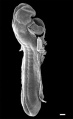
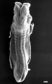
Human Embryo Carnegie stage 11
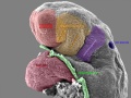
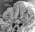
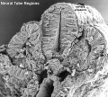
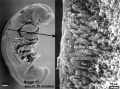
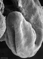
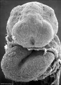
Week 4 Carnegie stage 11 25 days, 20 somite pairs. This is a scanning EM left ventrolateral view of the embryo head.
Features shown include the stomodeum, buccopharyngeal membrane, brain folds, first pharyngeal arch and heart tube.
labeled overlay image version and bright field image version of this image are also available.
- Stage 11 SEM Images: dorsolateral whole embryo | dorsal embryo | lateral embryo | lateral head | lateral head with overlay | embryo cross-section | ventrolateral head | ventrolateral head with overlay | ventral head | buccopharyngeal membrane | neural crest | posterior neuropore | anterior neuropore | Carnegie stage 11
- Human Embryo (stage 11)
dorsolateral whole embryo
dorsal embryo
lateral embryo
lateral head
lateral head with overlay

embryo cross-section
embryo cross-section label
neural cross-section label

ventrolateral head
ventrolateral head with overlay

ventral head

buccopharyngeal membrane
neural crest
posterior neuropore
anterior neuropore


Image Source: Scanning electron micrographs of the Carnegie stages of the early human embryos are reproduced with the permission of Prof Kathy Sulik, from embryos collected by Dr. Vekemans and Tania Attié-Bitach. Images are for educational purposes only and cannot be reproduced electronically or in writing without permission.
- Carnegie Stages: 1 | 2 | 3 | 4 | 5 | 6 | 7 | 8 | 9 | 10 | 11 | 12 | 13 | 14 | 15 | 16 | 17 | 18 | 19 | 20 | 21 | 22 | 23 | About Stages | Timeline
Image version links: Large 1000px | 800px | Medium 600px | Small 400px
Cite this page: Hill, M.A. (2024, April 19) Embryology Stage11 sem3c.jpg. Retrieved from https://embryology.med.unsw.edu.au/embryology/index.php/File:Stage11_sem3c.jpg
- © Dr Mark Hill 2024, UNSW Embryology ISBN: 978 0 7334 2609 4 - UNSW CRICOS Provider Code No. 00098G
File history
Click on a date/time to view the file as it appeared at that time.
| Date/Time | Thumbnail | Dimensions | User | Comment | |
|---|---|---|---|---|---|
| current | 18:12, 7 September 2009 |  | 375 × 400 (16 KB) | S8600021 (talk | contribs) |
You cannot overwrite this file.
File usage
The following 6 pages use this file: