File:Stage11 bf1.jpg
Original file (429 × 1,000 pixels, file size: 42 KB, MIME type: image/jpeg)
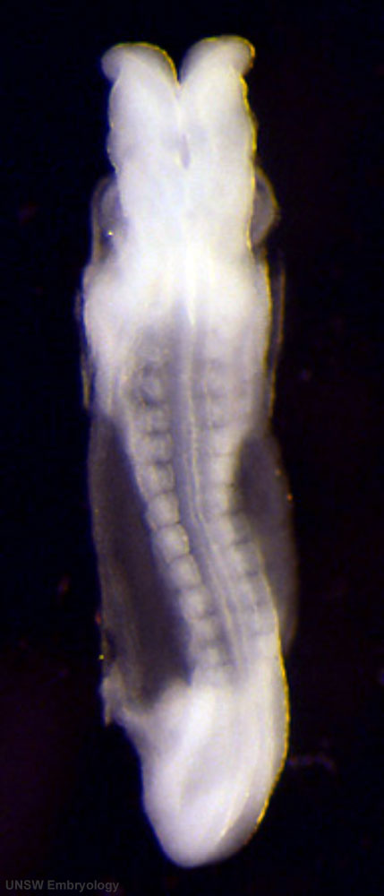
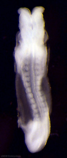
Human Embryo Carnegie stage 11
Carnegie stage 11, 24 days, 13 somite pairs
Facts: Week 4, 23 - 26 days, 2.5 - 4.5 mm, Somite Number 13 - 20
View: This is a dorsal view of embryo.
Features: neural tube, brain folds, somite pairs, heart, amnion
Scanning EM image version of whole embryo is also available.
- Stage 11 Images: BF1 - dorsal view | BF2 - lateral view | BF3 - ventral with scale bar | BF4 - ventral view | BF5 - lateral view | BF6 - ventral view | BF7 - Kyoto embryo | BF8 - ventral head | BF9 - ventral head | BF10 - dorsal neural | BF11 - ventral embryo and yolk sac | Scanning EM embryo | Carnegie stage 11
Image Source: Scanning electron micrographs of the Carnegie stages of the early human embryos are reproduced with the permission of Prof Kathy Sulik, from embryos collected by Dr. Vekemans and Tania Attié-Bitach. Images are for educational purposes only and cannot be reproduced electronically or in writing without permission.
Template:Carnegie stages table 1
Image version links | Large 1000px | 800px | Medium 600px | Small 400px
Cite this page: Hill, M.A. (2024, April 16) Embryology Stage11 bf1.jpg. Retrieved from https://embryology.med.unsw.edu.au/embryology/index.php/File:Stage11_bf1.jpg
- © Dr Mark Hill 2024, UNSW Embryology ISBN: 978 0 7334 2609 4 - UNSW CRICOS Provider Code No. 00098G
File history
Click on a date/time to view the file as it appeared at that time.
| Date/Time | Thumbnail | Dimensions | User | Comment | |
|---|---|---|---|---|---|
| current | 14:05, 7 September 2009 | | 429 × 1,000 (42 KB) | S8600021 (talk | contribs) | Stage11day24somite13-dorsal-bf1.jpg |
You cannot overwrite this file.
File usage
The following 3 pages use this file: