File:Stage10 sem9.jpg

Original file (740 × 1,000 pixels, file size: 72 KB, MIME type: image/jpeg)
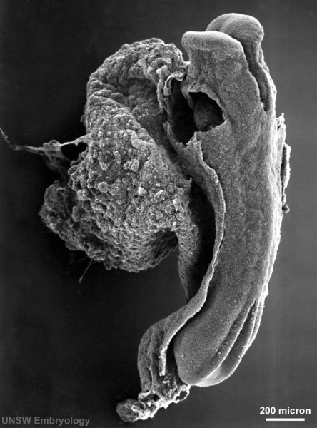
Human Embryo
Carnegie stage 10, 23 day, 11 somite pairs
Scanning EM dorsolateral view, yolk sac attached, amnion cut, section removed to view developing heart
scale bar 100 microns
Original File Name: Stage10day23somite11-lateral-sem1-rotated90.jpg
Stage10 sem9.jpg
Image version links: Large 1000px | 800px | Medium 600px | Small 400px
Horizontal Version: Large 1000px | 800px | Medium 600px | Small 400px
Image Source: Scanning electron micrographs of the Carnegie stages of the early human embryos are reproduced with the permission of Prof Kathy Sulik, from embryos collected by Dr. Vekemans and Tania Attié-Bitach. Images are for educational purposes only and cannot be reproduced electronically or in writing without permission.
Cite this page: Hill, M.A. (2024, April 24) Embryology Stage10 sem9.jpg. Retrieved from https://embryology.med.unsw.edu.au/embryology/index.php/File:Stage10_sem9.jpg
- © Dr Mark Hill 2024, UNSW Embryology ISBN: 978 0 7334 2609 4 - UNSW CRICOS Provider Code No. 00098G
File history
Click on a date/time to view the file as it appeared at that time.
| Date/Time | Thumbnail | Dimensions | User | Comment | |
|---|---|---|---|---|---|
| current | 19:03, 3 September 2009 | | 740 × 1,000 (72 KB) | S8600021 (talk | contribs) | Human Embryo Carnegie stage 10, 23 day, 11 somite pairs Scanning EM dorsolateral view, yolk sac attached, amnion cut, section removed to view developing heart scale bar 100 microns Original File Name: Stage10day23somite11-lateral-sem1-rotated90.jpg S |
You cannot overwrite this file.