File:Stage10 sem9.jpg: Difference between revisions
No edit summary |
No edit summary |
||
| (4 intermediate revisions by the same user not shown) | |||
| Line 1: | Line 1: | ||
Human Embryo | ==Human Embryo (Carnegie stage 10)== | ||
Carnegie stage 10, 23 day, 11 somite pairs | Carnegie stage 10, 23 day, 11 somite pairs | ||
Scanning EM dorsolateral view | ===Scanning EM dorsolateral view=== | ||
* scale bar | * scale bar 200 microns | ||
* yolk sac attached, amnion cut, connecting stalk (right) | * yolk sac attached, amnion cut, connecting stalk (right) | ||
* section removed to view developing heart in pericardial cavity | * section removed to view developing heart in pericardial cavity | ||
* open anterior and posterior neuropore | * open anterior and posterior neuropore | ||
* note appearance of extra-embryonic mesoderm covering yolk sac | * note appearance of extra-embryonic mesoderm covering yolk sac (bottom) | ||
* thin amniotic membrane (cut edges) | * thin amniotic membrane (cut edges) | ||
A vertical version of this image also available. | |||
Original File Name: Stage10day23somite11-lateral-sem1.jpg | |||
'''Image version links:''' [[:File:Stage10 sem9.jpg|Large 1000px]] | [[:File:Stage10 sem9a.jpg| 800px]] | | :'''Links:''' [[Carnegie stage 10]] | [[Week 4]] | [[:Category:Carnegie_Stage_10|Category:Carnegie Stage 10]] | [[Neural System Development]] | [[Cardiovascular System Development]] | [[Placenta Development]] | ||
===Image Links=== | |||
'''Image version links:''' [[:File:Stage10 sem10.jpg|Large 1000px]] | [[:File:Stage10 sem10a.jpg| 800px]] | | |||
[[:File:Stage10 sem10b.jpg|Medium 600px]] | [[:File:Stage10 sem10c.jpg|Small 400px]] | |||
'''Vertical version links:''' [[:File:Stage10 sem9.jpg|Large 1000px]] | [[:File:Stage10 sem9a.jpg| 800px]] | | |||
[[:File:Stage10 sem9b.jpg|Medium 600px]] | [[:File:Stage10 sem9c.jpg|Small 400px]] | [[:File:Stage10 sem9b.jpg|Medium 600px]] | [[:File:Stage10 sem9c.jpg|Small 400px]] | ||
{{Template:SEM}} | {{Template:SEM}} | ||
{{Template:Carnegie_stages}} | |||
{{Template:Footer}} | {{Template:Footer}} | ||
[[Category:Carnegie Stage 10]] [[Category:Week 4]] [[Category:Neural]] [[Category:Yolk Sac]] | |||
Latest revision as of 12:03, 17 March 2011
Human Embryo (Carnegie stage 10)
Carnegie stage 10, 23 day, 11 somite pairs
Scanning EM dorsolateral view
- scale bar 200 microns
- yolk sac attached, amnion cut, connecting stalk (right)
- section removed to view developing heart in pericardial cavity
- open anterior and posterior neuropore
- note appearance of extra-embryonic mesoderm covering yolk sac (bottom)
- thin amniotic membrane (cut edges)
A vertical version of this image also available.
Original File Name: Stage10day23somite11-lateral-sem1.jpg
- Links: Carnegie stage 10 | Week 4 | Category:Carnegie Stage 10 | Neural System Development | Cardiovascular System Development | Placenta Development
Image Links
Image version links: Large 1000px | 800px | Medium 600px | Small 400px
Vertical version links: Large 1000px | 800px |
Medium 600px | Small 400px
Image Source: Scanning electron micrographs of the Carnegie stages of the early human embryos are reproduced with the permission of Prof Kathy Sulik, from embryos collected by Dr. Vekemans and Tania Attié-Bitach. Images are for educational purposes only and cannot be reproduced electronically or in writing without permission.
- Carnegie Stages: 1 | 2 | 3 | 4 | 5 | 6 | 7 | 8 | 9 | 10 | 11 | 12 | 13 | 14 | 15 | 16 | 17 | 18 | 19 | 20 | 21 | 22 | 23 | About Stages | Timeline
Cite this page: Hill, M.A. (2024, April 18) Embryology Stage10 sem9.jpg. Retrieved from https://embryology.med.unsw.edu.au/embryology/index.php/File:Stage10_sem9.jpg
- © Dr Mark Hill 2024, UNSW Embryology ISBN: 978 0 7334 2609 4 - UNSW CRICOS Provider Code No. 00098G
File history
Click on a date/time to view the file as it appeared at that time.
| Date/Time | Thumbnail | Dimensions | User | Comment | |
|---|---|---|---|---|---|
| current | 19:03, 3 September 2009 | 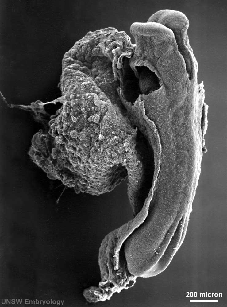 | 740 × 1,000 (72 KB) | S8600021 (talk | contribs) | Human Embryo Carnegie stage 10, 23 day, 11 somite pairs Scanning EM dorsolateral view, yolk sac attached, amnion cut, section removed to view developing heart scale bar 100 microns Original File Name: Stage10day23somite11-lateral-sem1-rotated90.jpg S |
You cannot overwrite this file.