File:Stage10 sem2.jpg: Difference between revisions
From Embryology
No edit summary |
No edit summary |
||
| (One intermediate revision by the same user not shown) | |||
| Line 1: | Line 1: | ||
==Human Embryo (Carnegie Stage 10)== | ==Human Embryo (Carnegie Stage 10)== | ||
* Scanning electron micrograph dorsolateral view of Carnegie Stage 10 embryo. | * Scanning electron micrograph dorsolateral view of Carnegie Stage 10 embryo. | ||
* '''Neural groove''' forming from neural plate on upper surface. | * '''Neural groove''' forming from neural plate on upper surface. | ||
** large brain fold region to left of image. | |||
** narrow spinal cord region to right of image. | |||
* '''Heart bulge''' can be seen on lower ventral surface. | * '''Heart bulge''' can be seen on lower ventral surface. | ||
* '''Connecting stalk''' to the right of image. | * '''Connecting stalk''' to the right of image. | ||
* '''Amniotic membrane''' cut edge shown. | * '''Amniotic membrane''' cut edge shown at edge of developing embryo. | ||
:'''Links:''' [[Carnegie stage 10]] | [[Lecture - Ectoderm Development|Lecture - Early Neural]] | [[Neural System Development]] | [[Week 4]] | :'''Links:''' [[:File:Stage10_sem6.jpg|vertical view]] | [[:File:Stage10_sem2.jpg|horizontal view]] | [[Carnegie stage 10]] | [[Lecture - Ectoderm Development|Lecture - Early Neural]] | [[Neural System Development]] | [[Week 4]] | ||
Latest revision as of 10:39, 22 November 2011
Human Embryo (Carnegie Stage 10)
- Scanning electron micrograph dorsolateral view of Carnegie Stage 10 embryo.
- Neural groove forming from neural plate on upper surface.
- large brain fold region to left of image.
- narrow spinal cord region to right of image.
- Heart bulge can be seen on lower ventral surface.
- Connecting stalk to the right of image.
- Amniotic membrane cut edge shown at edge of developing embryo.
- Links: vertical view | horizontal view | Carnegie stage 10 | Lecture - Early Neural | Neural System Development | Week 4
Image version links: Large 1000px | 800px |
Medium 600px | Small 400px
Image Source: Scanning electron micrographs of the Carnegie stages of the early human embryos are reproduced with the permission of Prof Kathy Sulik, from embryos collected by Dr. Vekemans and Tania Attié-Bitach. Images are for educational purposes only and cannot be reproduced electronically or in writing without permission.
- Carnegie Stages: 1 | 2 | 3 | 4 | 5 | 6 | 7 | 8 | 9 | 10 | 11 | 12 | 13 | 14 | 15 | 16 | 17 | 18 | 19 | 20 | 21 | 22 | 23 | About Stages | Timeline
Cite this page: Hill, M.A. (2024, April 24) Embryology Stage10 sem2.jpg. Retrieved from https://embryology.med.unsw.edu.au/embryology/index.php/File:Stage10_sem2.jpg
- © Dr Mark Hill 2024, UNSW Embryology ISBN: 978 0 7334 2609 4 - UNSW CRICOS Provider Code No. 00098G
File history
Click on a date/time to view the file as it appeared at that time.
| Date/Time | Thumbnail | Dimensions | User | Comment | |
|---|---|---|---|---|---|
| current | 12:27, 23 August 2009 | 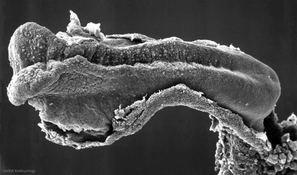 | 1,000 × 590 (60 KB) | S8600021 (talk | contribs) | Stage10day21somite4-5dorsolateralsem2v2.jpg |
You cannot overwrite this file.
File usage
The following 14 pages use this file:
- 2010 BGD Lecture - Development of the Embryo/Fetus 1
- 2010 BGD Practical 6 - Week 4
- 2010 Lab 3
- 2011 Lab 3 - Week 4
- ANAT3411 Neuroanatomy
- BGDA Lecture - Development of the Embryo/Fetus 1
- BGDA Lecture - Development of the Nervous System
- BGDA Practical 7 - Week 4
- Carnegie stage 10
- Carnegie stage 10 gallery
- Embryonic Development
- Human Embryo - Scanning electron microscopy
- Human Embryo SEM
- Talk:2011 Lab 3