File:Stage10 sem1.jpg: Difference between revisions
From Embryology
mNo edit summary |
|||
| Line 1: | Line 1: | ||
==Human Embryo SEM (Carnegie stage 10)== | ==Human Embryo SEM (Carnegie stage 10)== | ||
[[Carnegie stage 10]], [[Week 3]] (21 days, 4 somite pairs) {{GA}} week 5. Ventral view. | |||
{| | {| width=800px| | ||
| | | width=200px|200 | ||
| width=200px|200 | |||
| width=400px|400 | |||
| width=200px|200 | |||
|} | |} | ||
Revision as of 11:40, 4 May 2015
Human Embryo SEM (Carnegie stage 10)
Carnegie stage 10, Week 3 (21 days, 4 somite pairs) GA week 5. Ventral view.
| 200 | 200 | 400 | 200 |
Image Source: Scanning electron micrographs of the Carnegie stages of the early human embryos are reproduced with the permission of Prof Kathy Sulik, from embryos collected by Dr. Vekemans and Tania Attié-Bitach. Images are for educational purposes only and cannot be reproduced electronically or in writing without permission.
- Carnegie Stages: 1 | 2 | 3 | 4 | 5 | 6 | 7 | 8 | 9 | 10 | 11 | 12 | 13 | 14 | 15 | 16 | 17 | 18 | 19 | 20 | 21 | 22 | 23 | About Stages | Timeline
Cite this page: Hill, M.A. (2024, April 19) Embryology Stage10 sem1.jpg. Retrieved from https://embryology.med.unsw.edu.au/embryology/index.php/File:Stage10_sem1.jpg
- © Dr Mark Hill 2024, UNSW Embryology ISBN: 978 0 7334 2609 4 - UNSW CRICOS Provider Code No. 00098G
File history
Click on a date/time to view the file as it appeared at that time.
| Date/Time | Thumbnail | Dimensions | User | Comment | |
|---|---|---|---|---|---|
| current | 12:26, 23 August 2009 | 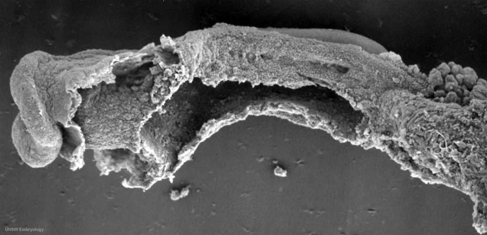 | 1,000 × 484 (46 KB) | S8600021 (talk | contribs) | Stage10day21somite4-5ventrolateralsem1v2.jpg |
You cannot overwrite this file.