File:Stage10 bf5c.jpg: Difference between revisions
From Embryology
No edit summary |
No edit summary |
||
| Line 1: | Line 1: | ||
Facts: Week 4, 22 - 23 days, 2 - 3.5 mm, Somite Number 4 - 12 | |||
View: This is a dorsal view of the embryo. Top embryo is an early stage 10, bottom is late stage 10. Amniotic membrane removed. | |||
Features: Somite Number 4 - 12, rostral neuropore, neural folds in region of developing brain, neural tube, somites, caudal neuropore, neural fold fuses, remnant of amniotic sac | |||
(note there are 2 separate images of this stage) | |||
Identify: rostral neuropore, neural folds in region of developing brain, neural tube, somites (note the different number formed), caudal neuropore, neural fold fuses, cut edge of amniotic sac | |||
Revision as of 14:58, 4 November 2009
Facts: Week 4, 22 - 23 days, 2 - 3.5 mm, Somite Number 4 - 12
View: This is a dorsal view of the embryo. Top embryo is an early stage 10, bottom is late stage 10. Amniotic membrane removed.
Features: Somite Number 4 - 12, rostral neuropore, neural folds in region of developing brain, neural tube, somites, caudal neuropore, neural fold fuses, remnant of amniotic sac
(note there are 2 separate images of this stage)
Identify: rostral neuropore, neural folds in region of developing brain, neural tube, somites (note the different number formed), caudal neuropore, neural fold fuses, cut edge of amniotic sac
File history
Click on a date/time to view the file as it appeared at that time.
| Date/Time | Thumbnail | Dimensions | User | Comment | |
|---|---|---|---|---|---|
| current | 14:52, 4 November 2009 | 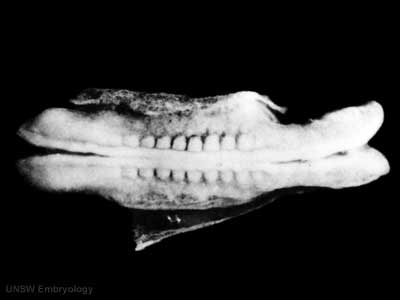 | 400 × 300 (7 KB) | S8600021 (talk | contribs) |
You cannot overwrite this file.
File usage
There are no pages that use this file.