File:Stage10 bf5b.jpg
Stage10_bf5b.jpg (600 × 450 pixels, file size: 23 KB, MIME type: image/jpeg)
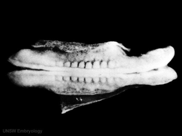
Human Embryo Carnegie Stage 10
Carnegie Stage 10 (22 - 23 days)
Facts: Week 4, 22 - 23 days, 2 - 3.5 mm, Somite Number 4 - 12
View: This is a dorsal view of the embryo. Top embryo is an early stage 10, bottom is late stage 10. Amniotic membrane removed.
Features: Somite Number 4 - 12, rostral neuropore, neural folds in region of developing brain, neural tube, somites, caudal neuropore, neural fold fuses, remnant of amniotic sac
(note there are 2 separate images of this stage)
Identify: rostral neuropore, neural folds in region of developing brain, neural tube, somites (note the different number formed), caudal neuropore, neural fold fuses, cut edge of amniotic sac Human Embryo
Original File name: Kyoto-stage-10.jpg
Image version links: Large 1000px | 800px | Medium 600px | Small 400px
Related Links: Carnegie stage 10 | original stage 10 page
- Carnegie Stages: 1 | 2 | 3 | 4 | 5 | 6 | 7 | 8 | 9 | 10 | 11 | 12 | 13 | 14 | 15 | 16 | 17 | 18 | 19 | 20 | 21 | 22 | 23 | About Stages | Timeline
Image source: The Kyoto Collection images are reproduced with the permission of Prof. Kohei Shiota and Prof. Shigehito Yamada, Anatomy and Developmental Biology, Kyoto University Graduate School of Medicine, Kyoto, Japan for educational purposes only and cannot be reproduced electronically or in writing without permission.
File history
Click on a date/time to view the file as it appeared at that time.
| Date/Time | Thumbnail | Dimensions | User | Comment | |
|---|---|---|---|---|---|
| current | 14:52, 4 November 2009 | | 600 × 450 (23 KB) | S8600021 (talk | contribs) |
You cannot overwrite this file.
File usage
The following 9 pages use this file: