File:Stage10 SEM1.jpg
Stage10_SEM1.jpg (277 × 450 pixels, file size: 28 KB, MIME type: image/jpeg)
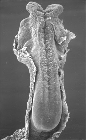
Carnegie Stages 10, 4-5 somites
MH - This description requires checking
Facts: Week 4, 22 - 23 days, 2 - 3.5 mm, Somite Number 4 - 12
View: This is a dorsal view of the embryo. Amniotic membrane removed.
Features: Somite Number 4 - 12, rostral neuropore, neural folds in region of developing brain, neural tube, somites, caudal neuropore, neural fold fuses, remnant of amniotic sac
Identify: rostral neuropore, neural folds in region of developing brain, neural tube, somites (note the different number formed), caudal neuropore, neural fold fuses, cut edge of amniotic sac
Events Ectoderm: Neural fold deeepens, edges approach midline, neural fold fuses, neural plate folds ventrally in brain region
Mesoderm: Somitogenesis, continued segmentation of paraxial mesoderm (4 - 12 somite pairs)
Image Source: Prof Kathy Sulik scanning electron micrographs of the Carnegie stages of the early human embryo. UNSW Embryology, no reproduction without permission. Carnegie Stages - Scanning Electron Micrography | Embryology page Created: 2007
Carnegie Stages Link
1 | 3 | 7 | 8 | 9 | 10 | 11 | 12 | 13 | 14 | 15 | 16 | 17 | 18 | 19 | 20 | 21 | 22 | 23
About Carnegie Stages
Carnegie stages are named after the famous US Institute which began collecting and classifying embryos in the early 1900's. Stages are based on the external and/or internal morphological development of the embryo, and are not directly dependent on either age or size. The human embryonic period proper is divided into 23 Carnegie stages. Carnegie stages are based on the external and/or internal morphological development of the embryo, and are not directly dependent on either age or size. Criteria beyond morphological features include age in days, number of somites present, and embryonic length.
The Kyoto Collection images are reproduced with the permission of Prof. Kohei Shiota. Scanning electron micrographs of the Carnegie stages of the early human embryos are reproduced with the permission of Prof Kathy Sulik. Images are for educational tutorial/revision purposes and cannot be reproduced electronically or in writing without permission.
UNSW Embryology Links
Glossary Links
A | B | C | D | E | F | G | H | I | J | K | L | M | N | O | P | Q | R | S | T | U | V | W | X | Y | Z
- Dr Mark Hill 2009, UNSW Embryology ISBN: 978 0 7334 2609 4 - UNSW CRICOS Provider Code No. 00098G
File history
Click on a date/time to view the file as it appeared at that time.
| Date/Time | Thumbnail | Dimensions | User | Comment | |
|---|---|---|---|---|---|
| current | 15:32, 10 August 2009 | | 277 × 450 (28 KB) | MarkHill (talk | contribs) | Carnegie Stages 10, 4-5 somites Features: brain fold, neural groove, amniotic sac, presomitic mesoderm, embryonic disc, primitive node, primative streak, primative groove, connecting stalk Facts: Week 3, 21 days, 4 - 5 somites, View: Dorsal view amnioti |
You cannot overwrite this file.
File usage
The following 11 pages use this file: