File:Spleen histology 08.jpg: Difference between revisions
No edit summary |
|||
| Line 2: | Line 2: | ||
* Human spleen, lymphoid tissue, white pulp, splenic corpuscle / lymph follicle, central artery, germinal center | * Human spleen, lymphoid tissue, white pulp, splenic corpuscle / lymph follicle, central artery, germinal center | ||
* Stain: | * Stain: {{HE}} | ||
Vascular: splenic artery - trabecular arteries - central arteries (arterioles) covered with periarteriolar lymphoid sheaths (PALS) | Vascular: splenic artery - trabecular arteries - central arteries (arterioles) covered with periarteriolar lymphoid sheaths (PALS) | ||
{{Spleen Vignette}} | |||
{{Spleen Histology}} | {{Spleen Histology}} | ||
Latest revision as of 10:01, 19 July 2019
Human Spleen Histology
- Human spleen, lymphoid tissue, white pulp, splenic corpuscle / lymph follicle, central artery, germinal center
- Stain: (Stain - Haematoxylin Eosin)
Vascular: splenic artery - trabecular arteries - central arteries (arterioles) covered with periarteriolar lymphoid sheaths (PALS)
| Historic Embryology |
  The term should not be confused with the renal structure, a Malpighian corpuscle (renal corpuscle). |
Links: Histology | Histology Stains | Blue Histology images copyright Lutz Slomianka 1998-2009. The literary and artistic works on the original Blue Histology website may be reproduced, adapted, published and distributed for non-commercial purposes. See also the page Histology Stains.
Cite this page: Hill, M.A. (2024, April 23) Embryology Spleen histology 08.jpg. Retrieved from https://embryology.med.unsw.edu.au/embryology/index.php/File:Spleen_histology_08.jpg
- © Dr Mark Hill 2024, UNSW Embryology ISBN: 978 0 7334 2609 4 - UNSW CRICOS Provider Code No. 00098G
Original file name: Spl20he.jpg
File history
Click on a date/time to view the file as it appeared at that time.
| Date/Time | Thumbnail | Dimensions | User | Comment | |
|---|---|---|---|---|---|
| current | 17:48, 21 February 2011 | 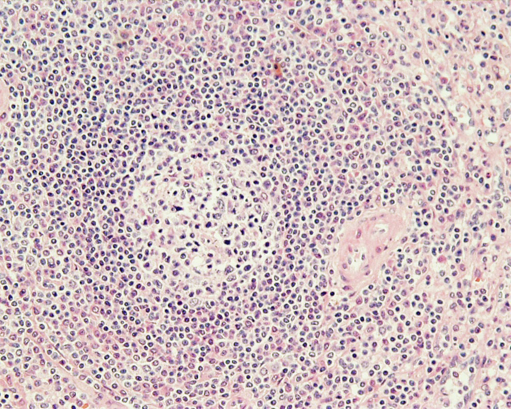 | 1,000 × 800 (304 KB) | S8600021 (talk | contribs) | ==Human Spleen== Lymphoid tissue, white pulp, splenic corpuscle / lymph follicle, central artery, germinal center Stain: H&E Original file name: Spl20he.jpg {{Blue Histology}} Category:Human Category:Spleen Category:Endocrine [[Category:H |
You cannot overwrite this file.
File usage
The following 11 pages use this file:
- ANAT2241 Lymphatic Tissue and Immune System
- Cardiovascular System - Spleen Development
- Histology
- Historic Embryology Vignette
- SH Practical - Lymphatic Quiz
- SH Practical - Lymphatic Structure and Organs
- Talk:SH Practical - Lymphatic Quiz
- File:Spleen histology 08.jpg
- Template:Spleen Histology gallery
- Template:Spleen Vignette
- Template talk:Spleen Histology