File:Spinal cord histology 03.jpg
Spinal_cord_histology_03.jpg (480 × 600 pixels, file size: 103 KB, MIME type: image/jpeg)
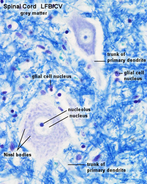
Spinal Cord Histology - Grey Matter
Most neurones have a light, large nucleus with a distinct nucleolus. The cytoplasm of many neurones contains fairly large amounts of rough endoplasmatic reticulum, which may aggregate within the cytoplasm of the neurone to form Nissl-bodies. Nissl-bodies are prominent in motor neurones located in the ventral horn of the grey matter of the spinal cord. The neurites are difficult to identify in most types of stained sections. Only the most proximal segments of the primary dendrites are seen clearly. The size of the perikaryon depends on the level of activity of the neurone and the length of the processes which the neurone has to support. An usable range for the size of the perikaryon would be 15 - 50 µm, although much smaller and much larger neuronal perikarya exist. (text Blue histology)
- Tissue - sheep spinal cord
- Stain - luxol fast blue/cresyl violet
- Spinal Cord: Overview 1 | Overview 2 | Overview animation | Grey matter | Grey matter | Grey matter | White matter | Overview unlabeled | Grey matter unlabeled 1 | Grey matter unlabeled 2 | White matter unlabeled 1 | Ependymal cells unlabeled
Links: Histology | Histology Stains | Blue Histology images copyright Lutz Slomianka 1998-2009. The literary and artistic works on the original Blue Histology website may be reproduced, adapted, published and distributed for non-commercial purposes. See also the page Histology Stains.
Cite this page: Hill, M.A. (2024, April 19) Embryology Spinal cord histology 03.jpg. Retrieved from https://embryology.med.unsw.edu.au/embryology/index.php/File:Spinal_cord_histology_03.jpg
- © Dr Mark Hill 2024, UNSW Embryology ISBN: 978 0 7334 2609 4 - UNSW CRICOS Provider Code No. 00098G
Spinal cord histology 03.jpg Original file name: spico041lf.jpg
File history
Click on a date/time to view the file as it appeared at that time.
| Date/Time | Thumbnail | Dimensions | User | Comment | |
|---|---|---|---|---|---|
| current | 10:16, 20 September 2012 | | 480 × 600 (103 KB) | Z8600021 (talk | contribs) | ==Spinal Cord Histology - Grey Matter== Most neurones have a light, large nucleus with a distinct nucleolus. The cytoplasm of many neurones contains fairly large amounts of rough endoplasmatic reticulum, which may aggregate within the cytoplasm of the ne |
You cannot overwrite this file.
File usage
The following 5 pages use this file: