File:Spaulding01.jpg: Difference between revisions
No edit summary |
No edit summary |
||
| Line 4: | Line 4: | ||
--[[User:S8600021|Mark Hill]] 16: | --[[User:S8600021|Mark Hill]] 16:36, March 26, 2011 (EST) CRL without knowing fixation shrinkage should be [[Carnegie stage 18]] or [[Carnegie stage 19]]. | ||
{{Spaulding1922}} | {{Spaulding1922}} | ||
Revision as of 16:51, 21 September 2011
Figure 1. Drawing of external genitalia of an embryo 16.8 mm. long
(Specimen No. 492), illustrating the genital-tubercle period. X 25.
--Mark Hill 16:36, March 26, 2011 (EST) CRL without knowing fixation shrinkage should be Carnegie stage 18 or Carnegie stage 19.
- Figure Links: Text | Text Figure 1 | Text Figure 2 | Plate 1 | Fig. 1 | Fig. 2 | Fig. 3 | Fig. 4 | Fig. 5 | Fig. 6 | Plate 2 | Fig. 7 | Fig. 8 | Fig. 9 | Fig. 10 | Fig. 11 | Fig. 12 | Fig. 13 | Fig. 14 | Fig. 15 | Fig. 16 | Fig. 17 | Fig. 18 | Fig. 19 | Fig. 20 | Fig. 21 | Fig. 22 | Plate 3 | Fig. 23 | Fig. 24 | Fig. 25 | Fig. 26 | Fig. 27 | Fig. 28 | Fig. 29 | Plate 4 | Fig. 30 | Fig. 31 | Fig. 32 |Fig. 33 | Fig. 34 | Fig. 35 | Fig. 36 | Fig. 37 | Fig. 38 | Fig. 39 | Fig. 40 | Fig. 41 | Fig. 42 | Fig. 43 | Fig. 44 | Fig. 45 | Fig. 46 | Fig. 47 | Fig. 48 | Fig. 49 | Fig. 50 | Fig. 51 | Fig. 52 | Fig. 53 | Fig. 54
| Historic Disclaimer - information about historic embryology pages |
|---|
 |
Reference
Spaulding MH. The development of the external genitalia in the human embryo. (1921) Contrib. Embryol., Carnegie Inst. Wash. Publ. 81, 13: 69 – 88.
Cite this page: Hill, M.A. (2024, April 18) Embryology Spaulding01.jpg. Retrieved from https://embryology.med.unsw.edu.au/embryology/index.php/File:Spaulding01.jpg
- © Dr Mark Hill 2024, UNSW Embryology ISBN: 978 0 7334 2609 4 - UNSW CRICOS Provider Code No. 00098G
| Historic Disclaimer - information about historic embryology pages |
|---|
|
Reference
The development of the external genitalia in the human embryo By Milo Herrick Spaulding, Of the University of Montmxa, Stale College of Agriculture, Bozeman. With four plates and two text-figures.
File history
Click on a date/time to view the file as it appeared at that time.
| Date/Time | Thumbnail | Dimensions | User | Comment | |
|---|---|---|---|---|---|
| current | 16:36, 26 March 2011 | 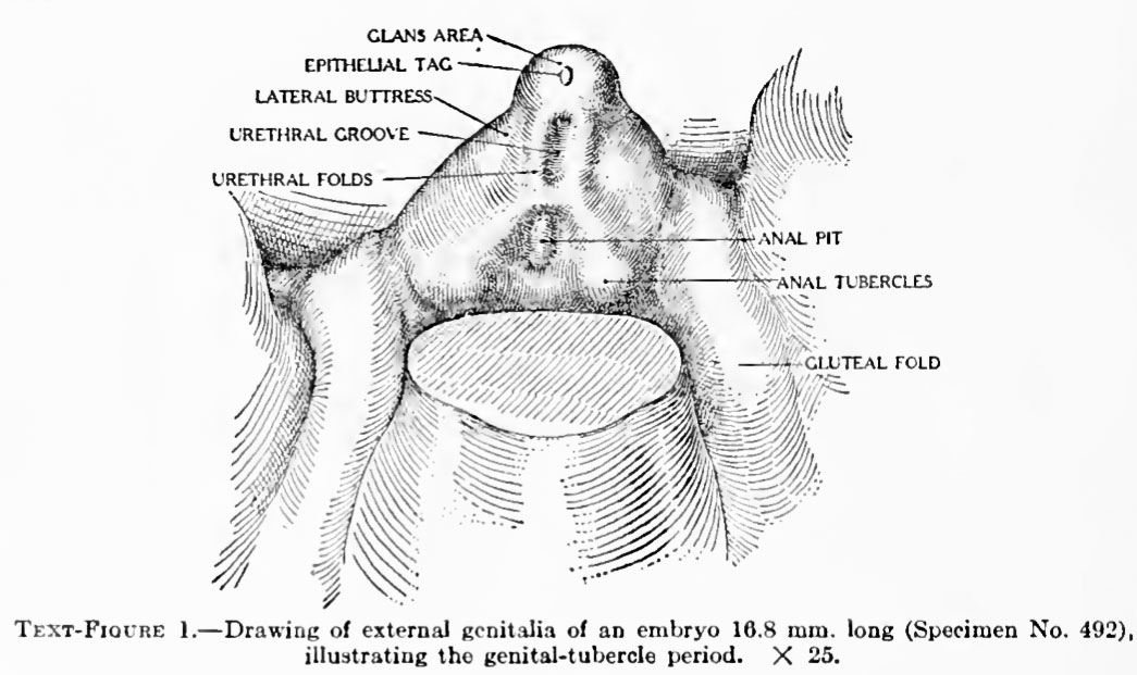 | 1,045 × 621 (111 KB) | S8600021 (talk | contribs) | ==Figure 1. Drawing of external genitalia of an embryo 16.8 mm. long== (Specimen No. 492), illustrating the genital-tubercle period. X 25. {{Template:Historic Disclaimer}} ==Reference== The development of the external genitalia in the human embryo By |
You cannot overwrite this file.
File usage
The following 3 pages use this file: