File:Spaulding-plate02.jpg: Difference between revisions
mNo edit summary |
mNo edit summary |
||
| Line 5: | Line 5: | ||
File:Spaulding-fig09.jpg|Fig. 9. Carnegie Embryo No. 28 | File:Spaulding-fig09.jpg|Fig. 9. Carnegie Embryo No. 28 | ||
File:Spaulding-fig10.jpg|Fig. 10. Carnegie Embryo No. 389a | File:Spaulding-fig10.jpg|Fig. 10. Carnegie Embryo No. 389a | ||
</gallery> | |||
<gallery> | |||
File:Spaulding-fig11.jpg|Fig. 11. Carnegie Embryo No. 2023 | File:Spaulding-fig11.jpg|Fig. 11. Carnegie Embryo No. 2023 | ||
File:Spaulding-fig12.jpg|Fig. 12. Carnegie Embryo No. 1750 | File:Spaulding-fig12.jpg|Fig. 12. Carnegie Embryo No. 1750 | ||
File:Spaulding-fig13.jpg|Fig. 13. Carnegie Embryo No. 684 | File:Spaulding-fig13.jpg|Fig. 13. Carnegie Embryo No. 684 | ||
File:Spaulding-fig14.jpg|Fig. 14. Carnegie Embryo No. 194 | File:Spaulding-fig14.jpg|Fig. 14. Carnegie Embryo No. 194 | ||
</gallery> | |||
<gallery> | |||
File:Spaulding-fig15.jpg|Fig. 15. Carnegie Embryo No.590 | File:Spaulding-fig15.jpg|Fig. 15. Carnegie Embryo No.590 | ||
File:Spaulding-fig16.jpg|Fig. 16. Carnegie Embryo No. 1900-60b | File:Spaulding-fig16.jpg|Fig. 16. Carnegie Embryo No. 1900-60b | ||
File:Spaulding-fig17.jpg|Fig. 17. Carnegie Embryo No.879c | File:Spaulding-fig17.jpg|Fig. 17. Carnegie Embryo No.879c | ||
File:Spaulding-fig18.jpg|Fig. 18. Carnegie Embryo No.1022d | File:Spaulding-fig18.jpg|Fig. 18. Carnegie Embryo No.1022d | ||
</gallery> | |||
<gallery> | |||
File:Spaulding-fig19.jpg|Fig. 19. Carnegie Embryo No. 2393 | File:Spaulding-fig19.jpg|Fig. 19. Carnegie Embryo No. 2393 | ||
File:Spaulding-fig20.jpg|Fig. 20. Carnegie Embryo No.1900-60a. | File:Spaulding-fig20.jpg|Fig. 20. Carnegie Embryo No.1900-60a. | ||
Revision as of 00:26, 15 April 2015
Plate 2
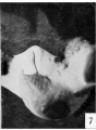
Fig. 7. Carnegie Embryo No. 1936
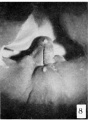
Fig. 8. Carnegie Embryo No. 955
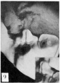
Fig. 9. Carnegie Embryo No. 28
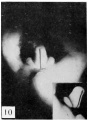
Fig. 10. Carnegie Embryo No. 389a


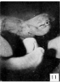
Fig. 11. Carnegie Embryo No. 2023
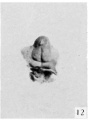
Fig. 12. Carnegie Embryo No. 1750
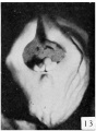
Fig. 13. Carnegie Embryo No. 684
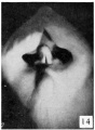
Fig. 14. Carnegie Embryo No. 194


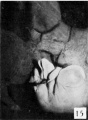
Fig. 15. Carnegie Embryo No.590
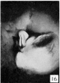
Fig. 16. Carnegie Embryo No. 1900-60b
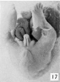
Fig. 17. Carnegie Embryo No.879c
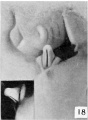
Fig. 18. Carnegie Embryo No.1022d


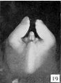
Fig. 19. Carnegie Embryo No. 2393
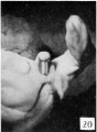
Fig. 20. Carnegie Embryo No.1900-60a.
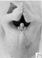
Fig. 21. Carnegie Embryo No. 950
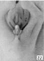
Fig. 22. Carnegie Embryo 1358h


Fig. 7. No. 1936, 14 mm., male. X 12.
Fig. 8. No. 955, 17 mm., male. X 12.
Fig. 9. No. 28, 19 mm., male. X 6. (Insert, lateral view.)
Fig. 10. No. 3S9a, 21 mm., male. X 6. (Insert, lateral view.)
Fig. 11. No. 202.S, 15 mm., female. X 12. 12. No. 1750, 17.2 mm., female. X
Fig. 12.
Fig. 13. No. 684, 20 mm., female. X 6.
Fig. 14. No. 194, 21 mm., female. X 6.
Fig. 15,
Fig. 16
Fig. 17, Fig. is
Fig. 19.
Fig. 20
Fig. 21
Fig. 22 , No. 590. 23 mm., male. X 6.
â– No. 1900-606, 25 mm., male. X 6. â– No. 879e, 29.6 mm., male. X 6. . No. 1022d, 32 mm., male. X 0. (Insert, lateral view.) No. 2393, 23 mm., female. X 6. No. 1900-60a, 25 mm., female. X 6. . No. 950, 29 mm., female. X 6. â– No. 1358h, 33 mm., female. X 6.
- Figure Links: Text | Text Figure 1 | Text Figure 2 | Plate 1 | Fig. 1 | Fig. 2 | Fig. 3 | Fig. 4 | Fig. 5 | Fig. 6 | Plate 2 | Fig. 7 | Fig. 8 | Fig. 9 | Fig. 10 | Fig. 11 | Fig. 12 | Fig. 13 | Fig. 14 | Fig. 15 | Fig. 16 | Fig. 17 | Fig. 18 | Fig. 19 | Fig. 20 | Fig. 21 | Fig. 22 | Plate 3 | Fig. 23 | Fig. 24 | Fig. 25 | Fig. 26 | Fig. 27 | Fig. 28 | Fig. 29 | Plate 4 | Fig. 30 | Fig. 31 | Fig. 32 |Fig. 33 | Fig. 34 | Fig. 35 | Fig. 36 | Fig. 37 | Fig. 38 | Fig. 39 | Fig. 40 | Fig. 41 | Fig. 42 | Fig. 43 | Fig. 44 | Fig. 45 | Fig. 46 | Fig. 47 | Fig. 48 | Fig. 49 | Fig. 50 | Fig. 51 | Fig. 52 | Fig. 53 | Fig. 54
| Historic Disclaimer - information about historic embryology pages |
|---|
 |
Reference
Spaulding MH. The development of the external genitalia in the human embryo. (1921) Contrib. Embryol., Carnegie Inst. Wash. Publ. 81, 13: 69 – 88.
Cite this page: Hill, M.A. (2024, April 24) Embryology Spaulding-plate02.jpg. Retrieved from https://embryology.med.unsw.edu.au/embryology/index.php/File:Spaulding-plate02.jpg
- © Dr Mark Hill 2024, UNSW Embryology ISBN: 978 0 7334 2609 4 - UNSW CRICOS Provider Code No. 00098G
| Historic Disclaimer - information about historic embryology pages |
|---|
|
Reference
The development of the external genitalia in the human embryo By Milo Herrick Spaulding, Of the University of Montmxa, Stale College of Agriculture, Bozeman. With four plates and two text-figures.
File history
Click on a date/time to view the file as it appeared at that time.
| Date/Time | Thumbnail | Dimensions | User | Comment | |
|---|---|---|---|---|---|
| current | 23:41, 14 April 2015 | 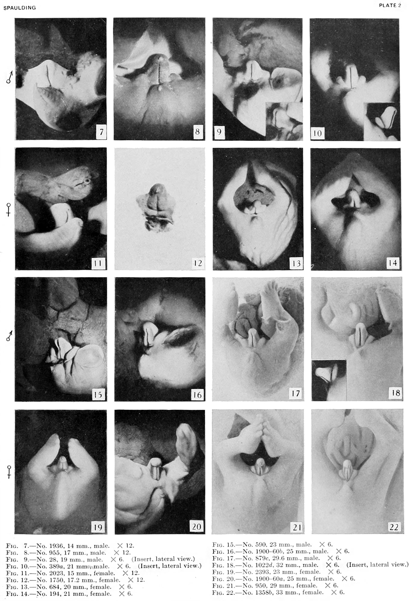 | 1,373 × 2,000 (449 KB) | Z8600021 (talk | contribs) | |
| 16:47, 26 March 2011 |  | 722 × 1,027 (119 KB) | S8600021 (talk | contribs) | ==Plate 2== {{Template:Historic Disclaimer}} ==Reference== The development of the external genitalia in the human embryo By Milo Herrick Spaulding, Of the University of Montmxa, Stale College of Agriculture, Bozeman. With four plates and two text-fi |
You cannot overwrite this file.
File usage
The following 2 pages use this file: