File:Spaulding-fig15.jpg: Difference between revisions
mNo edit summary |
mNo edit summary |
||
| (One intermediate revision by the same user not shown) | |||
| Line 1: | Line 1: | ||
==Fig. 15. Carnegie Embryo No.590== | ==Fig. 15. Carnegie Embryo No. 590== | ||
Carnegie Embryo No. {{CE590}} 23 mm., male. x 6. | |||
23 mm., | '''Stage 7''', 21 to 23 mm. CR ([[:File:Spaulding-fig10.jpg|fig. 10]], [[:File:Spaulding-fig15.jpg|fig. 15]], males; [[:File:Spaulding-fig14.jpg|fig. 14]], [[:File:Spaulding-fig19.jpg|fig. 19]], females). While there is a slight increase in the length of the phallus at this stage, the most conspicuous changes are the increase in the relative sex differences in the length of the urogenital opening and the greater development of the genital folds and the labio-scrotal swellings. In the males figured, the greater length of the urogenital opening is clearly seen to be in sharp contrast to the much shorter opening of the females. | ||
The urethral folds at this stage are more pronounced than in any of the embryos previously described. As a result of the deepening of the lateral depression between these folds and the cavernous portion of the shaft, their median portions now project from the shaft as compressed ridges. Distally they merge into the terminal glans area, while proximally they broaden out into the overhanging pre-anal enlargements (bulbo-urethral swellings?). As a result of this modification, the shaft of the phallus now shows clearly two distinct regions: the heavily thickened, cavernous portion, formed by the medial migration of the lateral buttresses, and the caudal urethral portion, represented by the projecting urethral folds outlining the urogenital opening, a condition which persists throughout the remainder of the phallus period. The labio-scrotal swellings have now grown into y-shaped enlargements, separated from the phallus by grooves more pronounced in the males than in the females. Cranially, these swellings are separated from each other by a triangular prolongation of the base of the phallus, so that there is no indication of the horseshoe shape which is so characteristic of most of the older illustrations. The caudal extremities of these swellings are joined together by a low, curving postanal ridge (slightly larger in the female), which is almost bisected by a median sagittal groove. | |||
{{ | {{Historic Disclaimer}} | ||
{{Spaulding1922}} | |||
{{ | |||
===Reference=== | ===Reference=== | ||
| Line 19: | Line 20: | ||
[[Category:Male]] | [[Category:Male]] | ||
[[Category:Carnegie Embryo 590]] | |||
Latest revision as of 14:01, 18 November 2017
Fig. 15. Carnegie Embryo No. 590
Carnegie Embryo No. 590 23 mm., male. x 6.
Stage 7, 21 to 23 mm. CR (fig. 10, fig. 15, males; fig. 14, fig. 19, females). While there is a slight increase in the length of the phallus at this stage, the most conspicuous changes are the increase in the relative sex differences in the length of the urogenital opening and the greater development of the genital folds and the labio-scrotal swellings. In the males figured, the greater length of the urogenital opening is clearly seen to be in sharp contrast to the much shorter opening of the females.
The urethral folds at this stage are more pronounced than in any of the embryos previously described. As a result of the deepening of the lateral depression between these folds and the cavernous portion of the shaft, their median portions now project from the shaft as compressed ridges. Distally they merge into the terminal glans area, while proximally they broaden out into the overhanging pre-anal enlargements (bulbo-urethral swellings?). As a result of this modification, the shaft of the phallus now shows clearly two distinct regions: the heavily thickened, cavernous portion, formed by the medial migration of the lateral buttresses, and the caudal urethral portion, represented by the projecting urethral folds outlining the urogenital opening, a condition which persists throughout the remainder of the phallus period. The labio-scrotal swellings have now grown into y-shaped enlargements, separated from the phallus by grooves more pronounced in the males than in the females. Cranially, these swellings are separated from each other by a triangular prolongation of the base of the phallus, so that there is no indication of the horseshoe shape which is so characteristic of most of the older illustrations. The caudal extremities of these swellings are joined together by a low, curving postanal ridge (slightly larger in the female), which is almost bisected by a median sagittal groove.
| Historic Disclaimer - information about historic embryology pages |
|---|
 |
- Figure Links: Text | Text Figure 1 | Text Figure 2 | Plate 1 | Fig. 1 | Fig. 2 | Fig. 3 | Fig. 4 | Fig. 5 | Fig. 6 | Plate 2 | Fig. 7 | Fig. 8 | Fig. 9 | Fig. 10 | Fig. 11 | Fig. 12 | Fig. 13 | Fig. 14 | Fig. 15 | Fig. 16 | Fig. 17 | Fig. 18 | Fig. 19 | Fig. 20 | Fig. 21 | Fig. 22 | Plate 3 | Fig. 23 | Fig. 24 | Fig. 25 | Fig. 26 | Fig. 27 | Fig. 28 | Fig. 29 | Plate 4 | Fig. 30 | Fig. 31 | Fig. 32 |Fig. 33 | Fig. 34 | Fig. 35 | Fig. 36 | Fig. 37 | Fig. 38 | Fig. 39 | Fig. 40 | Fig. 41 | Fig. 42 | Fig. 43 | Fig. 44 | Fig. 45 | Fig. 46 | Fig. 47 | Fig. 48 | Fig. 49 | Fig. 50 | Fig. 51 | Fig. 52 | Fig. 53 | Fig. 54
| Historic Disclaimer - information about historic embryology pages |
|---|
|
Reference
Spaulding MH. The development of the external genitalia in the human embryo. (1921) Contrib. Embryol., Carnegie Inst. Wash. Publ. 81, 13: 69 – 88.
Cite this page: Hill, M.A. (2024, April 19) Embryology Spaulding-fig15.jpg. Retrieved from https://embryology.med.unsw.edu.au/embryology/index.php/File:Spaulding-fig15.jpg
- © Dr Mark Hill 2024, UNSW Embryology ISBN: 978 0 7334 2609 4 - UNSW CRICOS Provider Code No. 00098G
Reference
Spaulding, M.H., The development of the external genitalia in the human embryo. Contributions to Embryology Carnegie Institution No.61 (1921). With four plates and two text-figures.
Cite this page: Hill, M.A. (2024, April 19) Embryology Spaulding-fig15.jpg. Retrieved from https://embryology.med.unsw.edu.au/embryology/index.php/File:Spaulding-fig15.jpg
- © Dr Mark Hill 2024, UNSW Embryology ISBN: 978 0 7334 2609 4 - UNSW CRICOS Provider Code No. 00098G
File history
Click on a date/time to view the file as it appeared at that time.
| Date/Time | Thumbnail | Dimensions | User | Comment | |
|---|---|---|---|---|---|
| current | 23:57, 14 April 2015 | 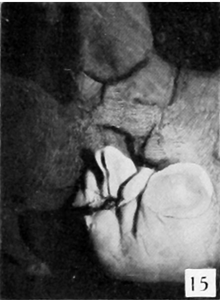 | 445 × 607 (38 KB) | Z8600021 (talk | contribs) |
You cannot overwrite this file.
File usage
The following 3 pages use this file: