File:Spaulding-fig02.jpg: Difference between revisions
mNo edit summary |
mNo edit summary |
||
| (One intermediate revision by the same user not shown) | |||
| Line 2: | Line 2: | ||
8 mm | 8 mm | ||
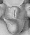
'''Stage 2''', 8 mm. ([[:File:Spaulding-fig02.jpg|fig. 2]], plate 1). The second stage, represented by the reconstruction model of an 8-mm. embryo, shows considerable advance in the development of the genital tubercle. This now forms a somewhat rounded mass which occupies almost the entire area between the umbilical cord and the base of the tail. Its cranial margin is but barely indicated by a slight groove between its apex and the umbilical cord. The apex is broadly rounded and from it the slightly convex caudal surface slopes toward the base of the tail. The caudal slope is almost bisected by the urethral groove, a shallow depression extending from the base to the tip of the tubercle. The basal end of the groove is separated from the anal pit by a narrow transverse bar. It is significant that, while there is this external separation of the primitive cloacal groove into the urethral groove and anal pit, the internal division of the cloacal cavity into urogenital sinus and rectum is not yet complete, at least in this embryo. The margins of the urethral groove are but slightly elevated into the urethral folds, although the margins of the anal pit are more pronounced. Laterally the caudal surface of the tubercle is rounded out into pronounced swellings. Almost in front of the tubercle the ventral body-wall bears a pair of swellings Iying in the umbilico-phallic angles, the significance of which has not yet been definitely ascertained. These masses show a gradual increase in size until the embryo reaches a length of 16 mm. They then apparently disappear in some embryos, while in others they undergo a caudal shifting; this would seem to indicate that they are the primordia of the labio-scrotal swellings, which definitely make their appearance in embryos 17 to 19 mm. long. As yet, however, a sufficient number of these younger embryos has not been examined to permit definite conclusions on this point. | |||
{{Historic Disclaimer}} | {{Historic Disclaimer}} | ||
| Line 14: | Line 17: | ||
{{Footer}} | {{Footer}} | ||
[[Category:Carnegie Embryo 792]] | |||
Latest revision as of 21:49, 21 April 2016
Fig. 2. Carnegie Embryo No. 792
8 mm
Stage 2, 8 mm. (fig. 2, plate 1). The second stage, represented by the reconstruction model of an 8-mm. embryo, shows considerable advance in the development of the genital tubercle. This now forms a somewhat rounded mass which occupies almost the entire area between the umbilical cord and the base of the tail. Its cranial margin is but barely indicated by a slight groove between its apex and the umbilical cord. The apex is broadly rounded and from it the slightly convex caudal surface slopes toward the base of the tail. The caudal slope is almost bisected by the urethral groove, a shallow depression extending from the base to the tip of the tubercle. The basal end of the groove is separated from the anal pit by a narrow transverse bar. It is significant that, while there is this external separation of the primitive cloacal groove into the urethral groove and anal pit, the internal division of the cloacal cavity into urogenital sinus and rectum is not yet complete, at least in this embryo. The margins of the urethral groove are but slightly elevated into the urethral folds, although the margins of the anal pit are more pronounced. Laterally the caudal surface of the tubercle is rounded out into pronounced swellings. Almost in front of the tubercle the ventral body-wall bears a pair of swellings Iying in the umbilico-phallic angles, the significance of which has not yet been definitely ascertained. These masses show a gradual increase in size until the embryo reaches a length of 16 mm. They then apparently disappear in some embryos, while in others they undergo a caudal shifting; this would seem to indicate that they are the primordia of the labio-scrotal swellings, which definitely make their appearance in embryos 17 to 19 mm. long. As yet, however, a sufficient number of these younger embryos has not been examined to permit definite conclusions on this point.
| Historic Disclaimer - information about historic embryology pages |
|---|
| Pages where the terms "Historic" (textbooks, papers, people, recommendations) appear on this site, and sections within pages where this disclaimer appears, indicate that the content and scientific understanding are specific to the time of publication. This means that while some scientific descriptions are still accurate, the terminology and interpretation of the developmental mechanisms reflect the understanding at the time of original publication and those of the preceding periods, these terms, interpretations and recommendations may not reflect our current scientific understanding. (More? Embryology History | Historic Embryology Papers) |
- Figure Links: Text | Text Figure 1 | Text Figure 2 | Plate 1 | Fig. 1 | Fig. 2 | Fig. 3 | Fig. 4 | Fig. 5 | Fig. 6 | Plate 2 | Fig. 7 | Fig. 8 | Fig. 9 | Fig. 10 | Fig. 11 | Fig. 12 | Fig. 13 | Fig. 14 | Fig. 15 | Fig. 16 | Fig. 17 | Fig. 18 | Fig. 19 | Fig. 20 | Fig. 21 | Fig. 22 | Plate 3 | Fig. 23 | Fig. 24 | Fig. 25 | Fig. 26 | Fig. 27 | Fig. 28 | Fig. 29 | Plate 4 | Fig. 30 | Fig. 31 | Fig. 32 |Fig. 33 | Fig. 34 | Fig. 35 | Fig. 36 | Fig. 37 | Fig. 38 | Fig. 39 | Fig. 40 | Fig. 41 | Fig. 42 | Fig. 43 | Fig. 44 | Fig. 45 | Fig. 46 | Fig. 47 | Fig. 48 | Fig. 49 | Fig. 50 | Fig. 51 | Fig. 52 | Fig. 53 | Fig. 54
| Historic Disclaimer - information about historic embryology pages |
|---|
| Pages where the terms "Historic" (textbooks, papers, people, recommendations) appear on this site, and sections within pages where this disclaimer appears, indicate that the content and scientific understanding are specific to the time of publication. This means that while some scientific descriptions are still accurate, the terminology and interpretation of the developmental mechanisms reflect the understanding at the time of original publication and those of the preceding periods, these terms, interpretations and recommendations may not reflect our current scientific understanding. (More? Embryology History | Historic Embryology Papers) |
Reference
Spaulding MH. The development of the external genitalia in the human embryo. (1921) Contrib. Embryol., Carnegie Inst. Wash. Publ. 81, 13: 69 – 88.
Cite this page: Hill, M.A. (2024, April 16) Embryology Spaulding-fig02.jpg. Retrieved from https://embryology.med.unsw.edu.au/embryology/index.php/File:Spaulding-fig02.jpg
- © Dr Mark Hill 2024, UNSW Embryology ISBN: 978 0 7334 2609 4 - UNSW CRICOS Provider Code No. 00098G
Reference
Spaulding, M.H., The development of the external genitalia in the human embryo. Contributions to Embryology Carnegie Institution No.61 (1921). With four plates and two text-figures.
Cite this page: Hill, M.A. (2024, April 16) Embryology Spaulding-fig02.jpg. Retrieved from https://embryology.med.unsw.edu.au/embryology/index.php/File:Spaulding-fig02.jpg
- © Dr Mark Hill 2024, UNSW Embryology ISBN: 978 0 7334 2609 4 - UNSW CRICOS Provider Code No. 00098G
File history
Click on a date/time to view the file as it appeared at that time.
| Date/Time | Thumbnail | Dimensions | User | Comment | |
|---|---|---|---|---|---|
| current | 23:22, 14 April 2015 |  | 646 × 748 (67 KB) | Z8600021 (talk | contribs) |
You cannot overwrite this file.
File usage
The following 2 pages use this file:
