File:Smooth muscle histology 002.jpg
Original file (600 × 750 pixels, file size: 112 KB, MIME type: image/jpeg)
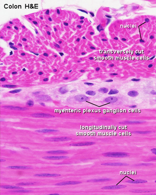
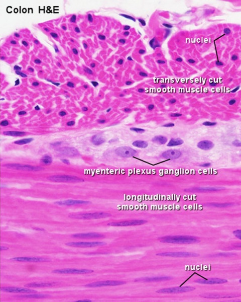
Smooth Muscle Histology
Histological section from the colon wall, stain H&E. Showing tunica muscularis and myenteric plexus.
From top to bottom of image:
- Outer longitudinal layer smooth muscle cells.
- Myenteric plexus (historic name Auerbach's plexus)
- Inner circular layer smooth muscle cells.
- Smooth Muscle Histology: Labeled Colon low | Labeled Colon high | Colon x40 | Colon x40 | Ileum x10 | Oesophagus x10 | Seminiferous tubule x40 | Uterus myometrium x10 | Uterus myometrium x40 |
Links: Histology | Histology Stains | Blue Histology images copyright Lutz Slomianka 1998-2009. The literary and artistic works on the original Blue Histology website may be reproduced, adapted, published and distributed for non-commercial purposes. See also the page Histology Stains.
Cite this page: Hill, M.A. (2024, April 18) Embryology Smooth muscle histology 002.jpg. Retrieved from https://embryology.med.unsw.edu.au/embryology/index.php/File:Smooth_muscle_histology_002.jpg
- © Dr Mark Hill 2024, UNSW Embryology ISBN: 978 0 7334 2609 4 - UNSW CRICOS Provider Code No. 00098G
File history
Click on a date/time to view the file as it appeared at that time.
| Date/Time | Thumbnail | Dimensions | User | Comment | |
|---|---|---|---|---|---|
| current | 01:46, 3 October 2011 | | 600 × 750 (112 KB) | S8600021 (talk | contribs) |
You cannot overwrite this file.