File:Smooth muscle histology 002.jpg: Difference between revisions
mNo edit summary |
|||
| Line 1: | Line 1: | ||
==Smooth Muscle Histology== | ==Smooth Muscle Histology== | ||
Histological section from the colon wall, | Histological section from the colon wall, {{HE}} | ||
Showing tunica muscularis and myenteric plexus (Auerbach's plexus). | |||
| Line 12: | Line 14: | ||
[[Category:Gastrointestinal Tract]] [[Category:Monkey]] | [[Category:Gastrointestinal Tract]] [[Category:Monkey]] [[Category:Neural Crest]] | ||
Revision as of 13:32, 1 May 2013
Smooth Muscle Histology
Histological section from the colon wall, (Stain - Haematoxylin Eosin)
Showing tunica muscularis and myenteric plexus (Auerbach's plexus).
Embryology Historic Terminology The historic name for myenteric plexus is Auerbach's plexus. In 1864 Auerbach first described the myenteric plexus lying between the longitudinal and circular muscle layers.
- Smooth Muscle Histology: Labeled Colon low | Labeled Colon high | Colon x40 | Colon x40 | Ileum x10 | Oesophagus x10 | Seminiferous tubule x40 | Uterus myometrium x10 | Uterus myometrium x40 |
Links: Histology | Histology Stains | Blue Histology images copyright Lutz Slomianka 1998-2009. The literary and artistic works on the original Blue Histology website may be reproduced, adapted, published and distributed for non-commercial purposes. See also the page Histology Stains.
Cite this page: Hill, M.A. (2024, April 25) Embryology Smooth muscle histology 002.jpg. Retrieved from https://embryology.med.unsw.edu.au/embryology/index.php/File:Smooth_muscle_histology_002.jpg
- © Dr Mark Hill 2024, UNSW Embryology ISBN: 978 0 7334 2609 4 - UNSW CRICOS Provider Code No. 00098G
File history
Click on a date/time to view the file as it appeared at that time.
| Date/Time | Thumbnail | Dimensions | User | Comment | |
|---|---|---|---|---|---|
| current | 01:46, 3 October 2011 | 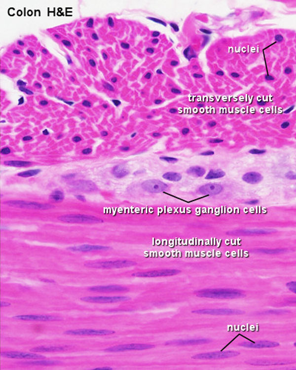 | 600 × 750 (112 KB) | S8600021 (talk | contribs) |
You cannot overwrite this file.