File:Small intestine villi and crypts.jpg
Small_intestine_villi_and_crypts.jpg (500 × 333 pixels, file size: 26 KB, MIME type: image/jpeg)
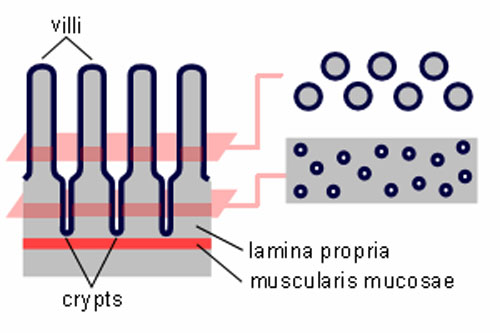
Intestinal Villi and Crypts of the Small Intestine
The entire intestinal mucosa forms intestinal villi (about one mm long), which increase the surface area by a factor of ~ ten. The surface of the villi is formed by a simple columnar epithelium. Each absorptive cell or enterocyte of the epithelium forms numerous microvilli (1 µm long and about 0.1 µm wide). Microvilli increase the surface area by a factor of ~ 20.
Between the intestinal villi we see the openings of simple tubular glands, the crypts of Lieberkühn. They extend through the lamina propria down to the muscularis mucosae. Undifferentiated cells close to the bottom of the crypts regenerate the epithelium (epithelial cell turnover time is less than one week). Other epithelial cells in the crypts correspond largely to those in the epithelium of the intestinal villi. One exception are Paneth cells which are located at the bottom of the crypts. They release a number of antibacterial substances, among them lysozyme, and are thought to be involved in the control of infections.
One function of the crypts of Lieberkühn is the secretion of "intestinal juice" (about 2 liter/day), which in its composition closely resembles extracellular fluid and which is rapidly reabsorbed. The only enzymes which can be demonstrated in the intestinal juice are enteropeptidase (or enterokinase), which activates the pancreatic enzyme trypsin, and small amounts of amylase. In addition to enterocytes, the epithelium is composed of mucus-secreting goblet cells and endocrine cells.
In addition to gastrin- and somatostatin-producing cells, we also find endocrine cells secreting cholecystokinin and secretin. Cholecystokinin stimulates the secretion of digestive enzymes in the pancreas and the contraction of the gall bladder. Secretin stimulates the pancreas to release "pancreatic juice", which is rich in bicarbonate ions. Secretin also amplifies the effects of cholecystokinin.
(Text from Blue Histology)
Links: Histology | Histology Stains | Blue Histology images copyright Lutz Slomianka 1998-2009. The literary and artistic works on the original Blue Histology website may be reproduced, adapted, published and distributed for non-commercial purposes. See also the page Histology Stains.
Cite this page: Hill, M.A. (2024, April 23) Embryology Small intestine villi and crypts.jpg. Retrieved from https://embryology.med.unsw.edu.au/embryology/index.php/File:Small_intestine_villi_and_crypts.jpg
- © Dr Mark Hill 2024, UNSW Embryology ISBN: 978 0 7334 2609 4 - UNSW CRICOS Provider Code No. 00098G
File history
Click on a date/time to view the file as it appeared at that time.
| Date/Time | Thumbnail | Dimensions | User | Comment | |
|---|---|---|---|---|---|
| current | 11:34, 15 April 2011 | | 500 × 333 (26 KB) | S8600021 (talk | contribs) | ==Intestinal Villi and Crypts of the Small Intestine== The entire intestinal mucosa forms intestinal villi (about one mm long), which increase the surface area by a factor of ~ ten. The surface of the villi is formed by a simple columnar epithelium. Each |
You cannot overwrite this file.
File usage
The following 3 pages use this file: