File:Skull CT abnormal 05.jpg
Original file (1,000 × 572 pixels, file size: 88 KB, MIME type: image/jpeg)
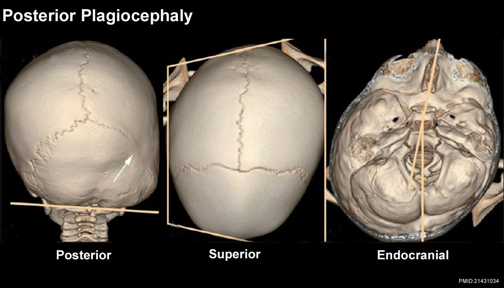
Posterior plagiocephaly
Posterior (A), superior (B) and endocranial (C) 3DCT volume rendered images show posterior plagiocephaly secondary to unilateral lambdoid fusion (arrow). The posterior skull base is tilted downward on the affected side (A), and the skull assumes a trapezoid shape (B). Note also that the posterior skull base axis (passing through the basion and opisthion) is rotated toward the side of lambdoid fusion and does not coincide with the anterior skull base axis (passing through the crista galli and basion) (C)
http://www.ijri.org/viewimage.asp?img=IndianJRadiolImaging_2011_21_1_49_76055_f7.jpg
File history
Click on a date/time to view the file as it appeared at that time.
| Date/Time | Thumbnail | Dimensions | User | Comment | |
|---|---|---|---|---|---|
| current | 16:59, 17 March 2012 | | 1,000 × 572 (88 KB) | Z8600021 (talk | contribs) |
You cannot overwrite this file.
File usage
The following 3 pages use this file: