File:Skin structure cartoon.jpg

Original file (700 × 720 pixels, file size: 52 KB, MIME type: image/jpeg)
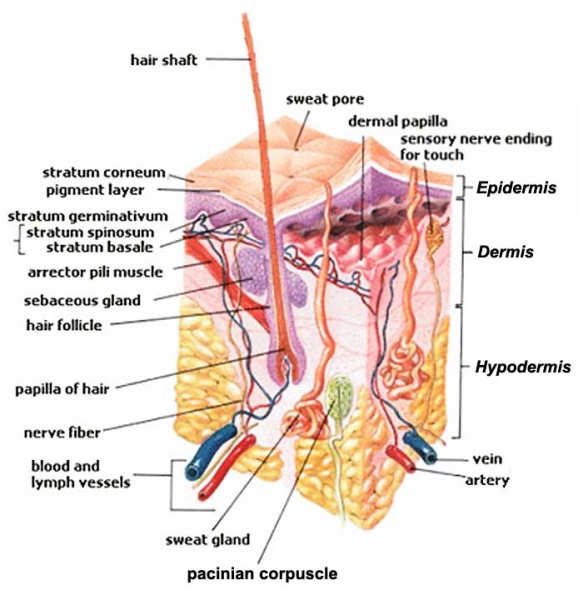
Skin Structure
Tissue combination of an epithelium (epidermis) and underlying connective tissue layers (dermis, hypodermis).
Features
- Basket cells - surround the base of hair follicles and can sense pressure. They are evaluated when assessing overall nerve health and condition.
- Blood vessels - carry nutrients and oxygen-rich blood to the cells that make up the layers of skin and carry away waste products.
- Hair erector muscle (arrector pili muscle) - tiny muscle connected to each hair follicle and the skin. When it contracts it causes the hair to stand erect, and a "goosebump" forms on the skin.
- Hair follicle - tube-shaped sheath that surrounds the part of the hair that is under the skin and nourishes the hair. It is located in the epidermis and the dermis.
- Hair shaft - the part of the hair that is above the skin.
- Langerhans Cells - cells attach themselves to antigens that invade damaged skin and alert the immune system to their presence.
- Melanocyte - cell that produces melanin, and is located in the basal layer of the epidermis.
- Merkel cells - tactile cells of neuroectodermal origin located in the basal layer of the epidermis.
- Pacinian corpuscle - (lamellar corpuscles) a nerve receptor located in the subcutaneous fatty tissue that responds to pressure and vibration.
- Rete pegs - (rete processes, rete ridges) epithelial extensions that project downward forming thickenings of the epidermis.
- Sebaceous gland - small, sack-shaped glands which release an oily substance onto the hair follicle that coats and protects the hair shaft from becoming brittle. These glands are located in the dermis.
- Sensory nerves - epidermis is innervated with sensory nerves. These nerves sense and transmit heat, pain, and other noxious sensations. When they are not functioning properly sensations such as numbness, pins-and-needles, pain, tingling, or burning may be felt. When evaluating a skin biopsy, total number, contiguity, diameter, branching, swelling, and overall health of the sensory nerves are assessed.
- Stratum corneum - outermost layer of the epidermis, and is comprised of dead skin cells. It protects the living cells beneath it by providing a tough barrier between the environment and the lower layers of the skin. The stratum corneum is useful for diagnosis because in some conditions it will become thinner than normal.
- Sweat gland - (sudoriferous gland) located in the epidermis and produce moisture (sweat) that is secreted through tiny ducts onto the surface of the skin (stratum corneum). When sweat evaporates, skin temperature is lowered.
Reference
Image Source: SEER Training Modules http://training.seer.cancer.gov/melanoma/anatomy/
Copyright
United States government creative works, including writing, images, and computer code, are usually prepared by officers or employees of the United States government as part of their official duties. A government work is generally not subject to copyright in the United States and there is generally no copyright restriction on reproduction, derivative works, distribution, performance, or display of a government work. Unless the work falls under an exception, anyone may, without restriction under U.S. copyright laws: Reproduce the work in print or digital form, Create derivative works, Perform the work publicly, Display the work, Distribute copies or digitally transfer the work to the public by sale or other transfer of ownership, or by rental, lease, or lending. https://www.usa.gov/government-works
Cite this page: Hill, M.A. (2024, April 25) Embryology Skin structure cartoon.jpg. Retrieved from https://embryology.med.unsw.edu.au/embryology/index.php/File:Skin_structure_cartoon.jpg
- © Dr Mark Hill 2024, UNSW Embryology ISBN: 978 0 7334 2609 4 - UNSW CRICOS Provider Code No. 00098G
File history
Click on a date/time to view the file as it appeared at that time.
| Date/Time | Thumbnail | Dimensions | User | Comment | |
|---|---|---|---|---|---|
| current | 16:31, 14 February 2012 | | 700 × 720 (52 KB) | S8600021 (talk | contribs) | |
| 18:25, 28 September 2009 |  | 396 × 407 (63 KB) | S8600021 (talk | contribs) | Image Source: SEER Training Modules http://training.seer.cancer.gov/melanoma/anatomy/ |
You cannot overwrite this file.
File usage
The following 14 pages use this file:
- 2009 Lecture 18
- 2010 Lecture 22
- A
- ANAT2241 Nervous Tissue
- ANAT2511 Basic Tissues
- ANAT2511 Integumentary System
- Foundations - Histology Cells and Tissues
- Foundations - Histology Epithelia and Skin
- H
- Human System Development
- Integumentary System - Hair Development
- Integumentary System Development
- Lecture - Integumentary Development
- P