File:Senior1919 fig10.jpg: Difference between revisions
(Z8600021 uploaded a new version of File:Senior1919 fig10.jpg) |
mNo edit summary |
||
| Line 10: | Line 10: | ||
{{Footer}} | {{Footer}} | ||
[[Category:Limb]] | |||
Latest revision as of 09:53, 12 August 2017
Fig. 10. Three reconstructions, each showing a sagittal segment (0.25 mm. thick) from the left leg of an embryo of the length indicated
Lateral aspect. X 19. A, 20 mm. ; B, 22 mm. ; C, 24.8 mm.
The tibia appears in all cases. P. indicates the position of the m. popliteus. c. m., a. communicans media; g, i. m., a. genu inferior medialis; g, m., a. genu media; i., a. interossea; {. m., interosseous membrane; p., a. poplitea; p. c, a. perforans cruris; p- d.^ a. tibialis anterior, pars distalis; p. p., a. poplitea profunda; p. s., a. poplitea superficialis; per., a. peronaea; r. a,, a. recurrens tibialis anterior; r. p., a. recurrens tibialis posterior; t, a., a. tibialis anterior; t, p. s., a. tibialis posterior superficialis.
Reference
Senior HD. The development of the arteries of the human lower extremity. (1919) Amer. J Anat. 22:1-11.
Cite this page: Hill, M.A. (2024, April 19) Embryology Senior1919 fig10.jpg. Retrieved from https://embryology.med.unsw.edu.au/embryology/index.php/File:Senior1919_fig10.jpg
- © Dr Mark Hill 2024, UNSW Embryology ISBN: 978 0 7334 2609 4 - UNSW CRICOS Provider Code No. 00098G
File history
Click on a date/time to view the file as it appeared at that time.
| Date/Time | Thumbnail | Dimensions | User | Comment | |
|---|---|---|---|---|---|
| current | 18:47, 31 October 2016 | 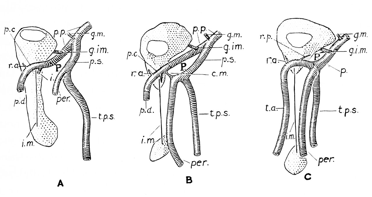 | 1,280 × 679 (134 KB) | Z8600021 (talk | contribs) | |
| 18:47, 31 October 2016 | 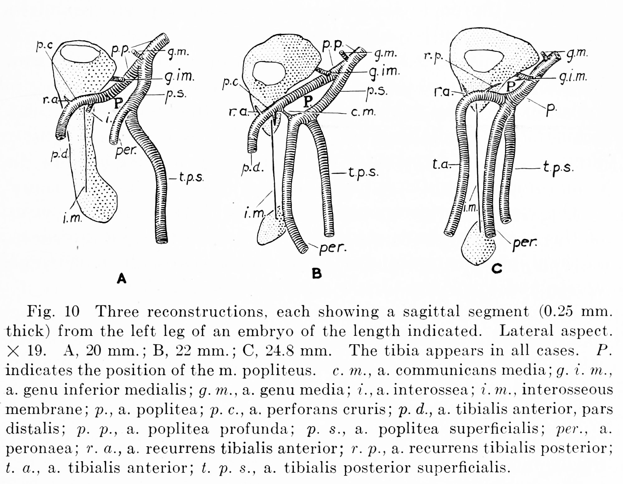 | 2,000 × 1,554 (424 KB) | Z8600021 (talk | contribs) | Fig. 10 Three reconstructions, each showing a sagittal segment (0.25 mm. thick) from the left leg of an embryo of the length indicated. Lateral aspect. X 19. A, 20 mm. ; B, 22 mm. ; C, 24.8 mm. The tibia appears in all cases. P. indicates the position... |
You cannot overwrite this file.
File usage
The following 2 pages use this file: