File:Senior1919 fig07.jpg

Original file (1,280 × 1,300 pixels, file size: 127 KB, MIME type: image/jpeg)
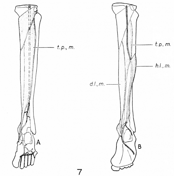
Fig, 7. Indicates the course of three arteries of the embryonic lower extremity
Represented diagrammatically as they would appear if persisting in the adult. The vessels which normally persist are indicated by shading.
A, A. poplitea profunda and a. interossea.
B. A. peronaea posterior superficialis. d. L m. flexor digitorum longus; h, L, m. flexor hallucis longus; L p., m. tibialis posterior.
Reference
Senior HD. The development of the arteries of the human lower extremity. (1919) Amer. J Anat. 22:1-11.
Cite this page: Hill, M.A. (2024, April 18) Embryology Senior1919 fig07.jpg. Retrieved from https://embryology.med.unsw.edu.au/embryology/index.php/File:Senior1919_fig07.jpg
- © Dr Mark Hill 2024, UNSW Embryology ISBN: 978 0 7334 2609 4 - UNSW CRICOS Provider Code No. 00098G
File history
Click on a date/time to view the file as it appeared at that time.
| Date/Time | Thumbnail | Dimensions | User | Comment | |
|---|---|---|---|---|---|
| current | 18:37, 31 October 2016 | | 1,280 × 1,300 (127 KB) | Z8600021 (talk | contribs) | |
| 18:37, 31 October 2016 |  | 2,000 × 1,888 (364 KB) | Z8600021 (talk | contribs) |
You cannot overwrite this file.
File usage
The following page uses this file: