File:Screen Shot 2017-10-05 at 3.29.59 pm.png
Screen_Shot_2017-10-05_at_3.29.59_pm.png (766 × 270 pixels, file size: 243 KB, MIME type: image/png)
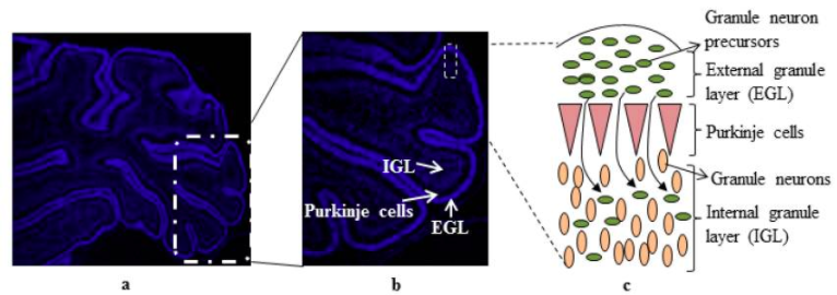
Cerebellum development of cells
A 7 day old rat cerebellum that has been sectioned to show the 3 layers of cells - external granule layer, purkinje cells and internal granular layer (a). Showing granule cell precursors proliferating to differentiate into granule neurons (b). Cell organisation (c) [1]
References
- ↑ <pubmed>4061865</pubmed>
Copyright
This article is an open access article distributed under the terms and conditions of the Creative Commons Attribution license You are free to:
Share — copy and redistribute the material in any medium or format Adapt — remix, transform, and build upon the material for any purpose, even commercially.
File history
Click on a date/time to view the file as it appeared at that time.
| Date/Time | Thumbnail | Dimensions | User | Comment | |
|---|---|---|---|---|---|
| current | 14:35, 5 October 2017 | 766 × 270 (243 KB) | Z5076158 (talk | contribs) | ==Cerebellum development of cells== A 7 day old rat cerebellum that has been sectioned to show the 3 layers of cells - external granule layer, purkinje cells and internal granular layer (a). Showing granule cell precursors proliferating to differentiat... |
You cannot overwrite this file.
File usage
The following 2 pages use this file: