File:Screen Shot 2017-10-05 at 3.29.59 pm.png
Screen_Shot_2017-10-05_at_3.29.59_pm.png (766 × 270 pixels, file size: 243 KB, MIME type: image/png)
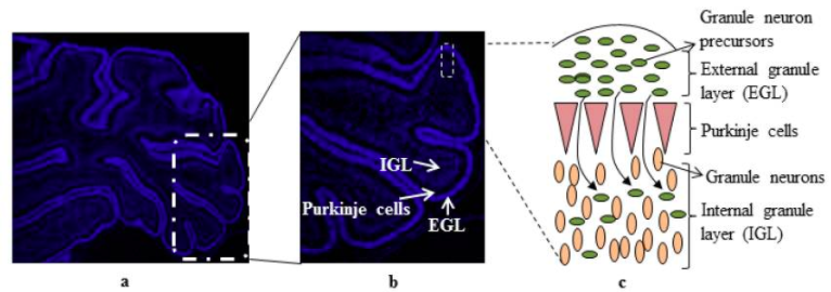
Cerebellum development of cells
A 7 day old rat cerebellum that has been sectioned to show the 3 layers of cells - external granule layer, purkinje cells and internal granular layer (a). Showing granule cell precursors proliferating to differentiate into granule neurons (b). Cell organisation (c) [1]
References
- ↑ <pubmed>4061865</pubmed>
Copyright
This article is an open access article distributed under the terms and conditions of the Creative Commons Attribution license You are free to:
Share — copy and redistribute the material in any medium or format Adapt — remix, transform, and build upon the material for any purpose, even commercially.
- Note - This image was originally uploaded as part of an undergraduate science student project and may contain inaccuracies in either description or acknowledgements. Students have been advised in writing concerning the reuse of content and may accidentally have misunderstood the original terms of use. If image reuse on this non-commercial educational site infringes your existing copyright, please contact the site editor for immediate removal.
File history
Click on a date/time to view the file as it appeared at that time.
| Date/Time | Thumbnail | Dimensions | User | Comment | |
|---|---|---|---|---|---|
| current | 14:35, 5 October 2017 | 766 × 270 (243 KB) | Z5076158 (talk | contribs) | ==Cerebellum development of cells== A 7 day old rat cerebellum that has been sectioned to show the 3 layers of cells - external granule layer, purkinje cells and internal granular layer (a). Showing granule cell precursors proliferating to differentiat... |
You cannot overwrite this file.
File usage
The following 2 pages use this file: