File:Schematic diagram of heart tube looping.png
From Embryology
No higher resolution available.
Schematic_diagram_of_heart_tube_looping.png (554 × 394 pixels, file size: 117 KB, MIME type: image/png)
Description
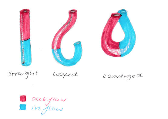
The heart tube begins as a straight tube, with blood entering caudally via the inflow and exiting cranially at the outflow tract. Heart tube looping occurs to the right, creating inflow and outflow portions of the tube. The inflow and outflow regions grow towards each other in the process of convergence, before separation creates the four-chambered heart.
References
Original student image based off: http://sheaheart.com/embryologyoftheheart/
- Note - This image was originally uploaded as part of an undergraduate science student project and may contain inaccuracies in either description or acknowledgements. Students have been advised in writing concerning the reuse of content and may accidentally have misunderstood the original terms of use. If image reuse on this non-commercial educational site infringes your existing copyright, please contact the site editor for immediate removal.
File history
Click on a date/time to view the file as it appeared at that time.
| Date/Time | Thumbnail | Dimensions | User | Comment | |
|---|---|---|---|---|---|
| current | 20:56, 4 October 2017 | | 554 × 394 (117 KB) | Z5076019 (talk | contribs) | Student Image based off [http://sheaheart.com/embryologyoftheheart/] |
You cannot overwrite this file.
File usage
The following 2 pages use this file: